A 28-year-old female patient was transported to our emergency department due to a recurrent episode of pneumonia of her right lung; fever, dyspnea and productive cough were the predominant symptoms. The woman was pregnant (in her last month of pregnancy) with no history of smoking, BMI 24,7 and the last three months she received antibiotic treatment twice, for right middle lobe pneumonia and right inferior lobe pneumonia, respectively.
Radiographic investigation revealed infiltrative lesions of the right lower lung and obliteration of the right costophrenic angle (Figure 1). Flexible bronchoscopy showed a significantly narrowed middle lobar bronchus, without any other endobronchial lesions (Figure 1). Expiratory/inspiratory CT scan demonstrated stenosis of middle lobar bronchus in expiration phase, with concomitant air entrapment in middle and inferior right lobe (Figure 2). Regarding her past medical history, it is worthy of note that she was submitted to an uneventful posterior spinal fusion with Harrington instrumentation, at the age of 14, due to severe idiopathic scoliosis (Cobb’s angle >40o) of the thoracic region and that she demonstrated similar episodes of pneumonia during the 8th and 9th month of her first pregnancy. The disease was efficiently controlled with antibiotic and bronchodilator treatment.
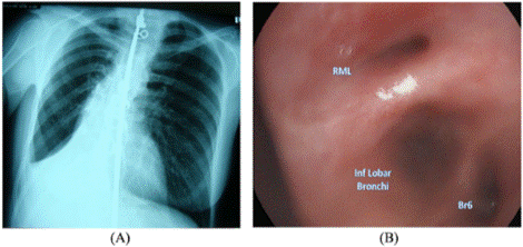
Figure 1. Chest X-ray (A) and bronchoscopic image (B). (A) Harrington rod, residual scoliosis of thoracic region, obliteration of right costophrenic angle and infiltrative lesions of right lower lung are illustrated. (B) Apparent stenosis of right middle lobar bronchus
Middle lobe syndrome, common variant immunodeficiency syndrome and recurrent pneumonia by atypical mycobacteria were excluded and we concluded that the narrowing of the right middle lobar bronchus was the result of pressure from the outside; presumably the abnormal shape of the thorax, due to the spinal fusion and residual scoliosis, and the concomitant rise of abdominal pressure, due to pregnancy. These conditions restricted the mobility of the thoracic cage, and in addition to the pressure of the lobar bronchus by the vertebral body (Figure 2), gave rise to atelectasis and pneumonia of the lower right lung.
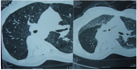
Figure 2. Inspiratory/Expiratory chest CT. The narrowing of the middle lobar bronchus caused by the vertebral body can be recognized (arrow)
A described effect of Harrington instrumentation is the flat back syndrome. In this patient, the flattening of the thoracic spine and the residual scoliosis, in combination with the pressure by the diaphragm, due to pregnancy, resulted in bronchial compression and narrowing. This, subsequently, led to restriction of lung function and recurrent infection of middle and lower right lung. The effects of posterior spinal fusion in pulmonary function have been widely reported in literature. Akazawa et al, in their study of 18 patients with adolescent idiopathic scoliosis (AIS) treated with posterior spinal fusion, reported restrictive ventilation defects in 27,7% of the patients 27 years after surgery [1]. Similar findings are reported by Upadhyay et al, who studied 35 patients with idiopathic scoliosis and observed post-operative reduction of vital capacity and significant increase of residual volume [2]. It seems possible from literature and from the experience with our patient that bronchial stenosis and reduced pulmonary function in cases of thoracic scoliosis can be the cause of respiratory insufficiency [3]. Undoubtedly, this hypothesis needs further investigation.
ST and EP contributed to patient management. ST, KD and KC drafted the initial manuscript. TK reviewed the manuscript. All authors contributed to writing the manuscript. All the authors have provided written consent for publication.
The authors have not declared a specific grant for this research from any funding agency in the public, commercial or not-for-profit sectors.
None declared.
Obtained.
Not commissioned; externally peer reviewed.
- Akazawa T, Kuroya S, Iinuma M (2018) Pulmonary function and thoracic deformities in adolescent idiopathic scoliosis 27 years or longer after spinal fusion with Harrington instrument. J Orthop Sci 23(1):45-50. [Crossref]
- Upadhyay SS, Day GA, Saji MJ (1993) Restrictive pattern of pulmonary functions in idiopathic and congenital scoliosis following spinal fusion. Eur Spine J 2(1):22-28.
- van Ooij A, van Belle A, Timmer R (2002) The destroyed lung syndrome: report of a case after Harrington rod instrumentation and fusion for idiopathic scoliosis. Spine (Phila Pa 1976) 27(14):337-341. [Crossref]