A 63-year-old non-smoker woman with a 15 years history of rheumatoid arthritis characterized by erosive poliarthrtitis, rheumatoid factor elevated titers, and positive testing for anti-CCP antibodies. The treatment included methotrexate, prednisolone and hydroxicloroquine. Despite of treatment, developed isquemic ulcer at left hand dorsum and nailfod infarction and digital ischemia of the left fingers [1].
Rheumatoid vasculitis is an inflammatory process that primarily affects small to medium-sized vessels. It’s highly heterogeneous clinically, with wide-spread organ involvement. The incidence has declined in the past several decades, but cutaneous remains the most common presentation. It tipically occurs in patients with long-standing erosive deforming Rheumatoid Arthritis, and its manifestation is heterogeneous, dependeing the size of the blood vessel. The skin can present purpura, nailfold infarcts, digital gangrene and cutaneous ulcers [2].
The photographs demonstrates a typical rheumatoid hand, with ulnar deviation, atrophy of interosseous muscles, metacarpophalangeal subluxation, and deformities (swan’s neck and boutonnière). there are two forms of rheumatoid vasculitis, represented by ulceration in the area of the third metacarpophalangeal joint and periungual infarction in the second one, as well as the discrete points of ischemia in the digital pulps. This image reflects the heterogeneity of the clinical presentations of this single entity [3].
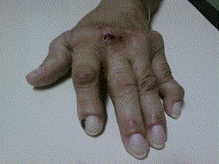
Figure 1. A typical rheumatoid hand, with ulnar deviation, atrophy of interosseous muscles, metacarpophalangeal subluxation, and deformities (swan’s neck and boutonnière).
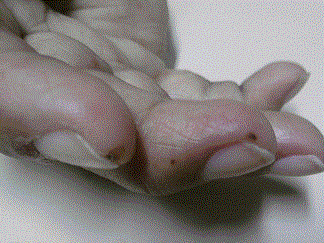
Figure 2. There are two forms of rheumatoid vasculitis, represented by ulceration in the third metacarpophalangeal joint and periungual infarction in the figure, as well as the discrete points of ischemia in the digital pulps.
- Makol A1, Matteson EL, Warrington KJ (2015) Rheumatoid vasculitis: an update. Curr Opin Rheumatol 27: 63-70. [crossref]
- Scott DG, Bacon PA, Tribe CR (1981) Systemic rheumatoid vasculitis: A clinical and laboratory study of 50 cases. Medicine (Baltimore) 60: 288-297.
- Makol A1, Crowson CS, Wetter DA, Sokumbi O, Matteson EL, et al. (2014) Vasculitis associated with rheumatoid arthritis: a case-control study. Rheumatology (Oxford) 53: 890-899. [crossref]
Editorial Information
Editor-in-Chief
Jose Luis Turabian
Complutense University, Madrid
Spain
Article Type
Case Report
2021 Copyright OAT. All rights reserv
Publication history
Received date: March 14, 2018
Accepted date: March 27, 2018
Published date: March 30, 2018
Copyright
© 2018 Paz OAG This is an open-access article distributed under the terms of the Creative Commons Attribution License, which permits unrestricted use, distribution, and reproduction in any medium, provided the original author and source are credited.
Citation
Paz OAG (2018) Rheumatoid vasculitis: same hand, heterogeneous clinical presentation. Trends Gen Pract 1: DOI: 10.15761/TGP.1000103
Corresponding author
Otávio Augusto Gomes Paz
University Center of Pará (CESUPA), Area of Environmental, Biological and Health Sciences. Belem, Brazil.
E-mail : bhuvaneswari.bibleraaj@uhsm.nhs.uk
Figure 1. A typical rheumatoid hand, with ulnar deviation, atrophy of interosseous muscles, metacarpophalangeal subluxation, and deformities (swan’s neck and boutonnière).
Figure 2. There are two forms of rheumatoid vasculitis, represented by ulceration in the third metacarpophalangeal joint and periungual infarction in the figure, as well as the discrete points of ischemia in the digital pulps.