Lung hypoplasia similarly to lung agenesis is a rare anomaly and frequently is accompanied by defects of other systems, usually cardiovascular. This makes complete straightforward diagnosis difficult. Complex congenital pulmonary and cardiovascular anomalies when suspected require extensive evaluation. First choice method for the evaluation of hemodynamic parameters, intracardiac defects is echocardiography. However, in cases with bronchopulmonary anomalies involving pulmonary circulation ultrasound with limited diagnostic window is insufficient. Most frequently the next step is radiological tomographic methods. Although nonionizing radiation makes magnetic resonance imaging (MRI) safer and more attractive especially in infants, however prolonged scan time, susceptibility to motion artefacts, intermediate spatial resolution and imperative general anesthesia precludes for fast, safe and informative investigation. Therefore, computed tomography (CT) with angiographic technique is the most frequently performed diagnostic modality for comprehensive evaluation of complex congenital thoracic pathology.
5-month-old male was referred to tertiary healthcare institution for poor weight gain, suspected cardiac anomalies and propensity to lung infections. Chest X-ray revealed right diaphragmatic hernia and malposition of the heart to the right side of the chest (Figure 1A). Blood test ruled out bacterial infection. Echocardiographic findings were consistent with subaortic ventricular septal defect, slight aortic dextraposition and pronounced pulmonary hypertension. Chest CT showed marked elevation of the right hemidiaphragm, most of the right hemithorax was occupied by the liver, no right lung atelectasis (Figure 1B). Abnormal right bronchial tree was detected with abrupt ending of the right upper and middle lobe bronchi. Right pulmonary artery was hypoplastic and supplied the right lower lobe, two right pulmonary veins entered liver tissue and coursed through liver parenchyma caudally to enter portal vein (Figures 1B, 1C and 1D). Smooth right border of the left atrium also was consistent with anomalous right pulmonary venous drainage. In addition, one of the right pulmonary artery branches was noticed to descend and traverse mediastinum to the left hemithorax, were hyperinflated parenchyma of the herniated right lung was observed (Figure 1B). Arterial phase of selective angiography showed filling of the hypoplastic right pulmonary artery with one of the branches traversing middle line of the body to the left hemithorax (Figure 1E), pulmonary venous phase was consistent with CT findings and depicted downward flow of contrast medium towards hepatic hilum (Figure 1F).
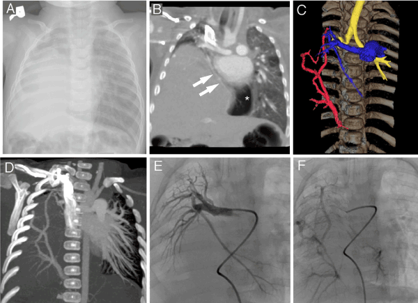
Figure 1.
A - frontal chest x-ray showing opaque right hemithorax
B - CT frontal maximum intensity projection reconstruction showing branch of the right pulmonary artery (two arrows) traversing mediastinum to supply the part of the right lung dislocated leftwards to left hemithorax (dislocated lung is of lower attenuation compared to left lung most probably because of hyperinflation - asterisk)
C - colored volume rendering image showing hypoplastic right pulmonary artery (blue), right pulmonary veins descending towards portal vein (red) and short trachea with high positioned carina (yellow)
D - maximum intensity projection showing right pulmonary vein drainage pattern
E - arterial phase of selective angiography showing right pulmonary artery
F - pulmonary venous phase of selective angiography showing right pulmonary veins
Infants suffering from complex bronchopulmonary and/or cardiovascular anomalies present with unspecific symptoms as pale or blue skin, rapid breathing, shortness of breath during feeding. Usually this precludes gain weight and requires more in-depth diagnostic imaging workup. More than decade existing non-invasive imaging methods proved to be safe and precise in making correct diagnosis in congenital thoracic bronchopulmonary and cardiovascular disease [1,2]. There are numerous cases with anomalous pulmonary vein return to portal system reported [3,4]. In our case dysplastic right lung, markedly elevated right hemidiaphragm and associated cardiovascular defects lead to unusual intrahepatic course of right pulmonary veins to portal system. Chest CT allowed precise description of all anatomic details associated with this complex bronchopulmonary and cardiovascular anomaly which were confirmed and visualized in a dynamic fashion using invasive selective angiography.
Compliance with Ethical Standards
Authors claim no funding received.
Authors declare that there is no actual or potential conflict of interest in relation to this article.
Parents of an infant were presented and signed Vilnius University Hospital informed consent forms.
- Raimondi F, Warin-Fresse K (2016) Computed tomography imaging in children with congenital heart disease: Indications and radiation dose optimization. Arch Cardiovasc Dis 109: 150-157. [Crossref]
- Sorensen C, Gach P, Pico H, Hugues N, Dabadie A, et al. (2016) Cardiac CT or MRI in pediatric practice: Which one to choose? Diagn Interv Imaging 97: 513-517. [Crossref]
- Dyer KT, Hlavacek AM, Meinel FG, De Cecco CN, McQuiston AD, et al. (2014) Imaging in congenital pulmonary vein anomalies: the role of computed tomography. Pediatr Radiol 44: 1158-1168; quiz 1155-1157. [Crossref]
- Aluja Jaramillo F, Hernandez C, Garzon JP, Sanchez Herrera AP, Velasco Morales ML (2017) Infracardiac type total anomalous pulmonary venous return with obstruction and dilatation of portal vein. Radiol Case Rep 12: 229-232. [Crossref]