Background: A 48 year old female with end stage renal disease underwent a living donor renal transplant. During the procedure she had a renal artery dissection necessitating reconstruction using a dacron graft.
Summary: Our patient had renal artery dissection during implantation for which the kidney had to be explanted and renal artery shortened to normal intima, leaving a 2 mm stump beyond hilum. Since there were no donor vessels available, we reconstructed the renal artery with a 6 mm dacron graft and reimplanted the kidney with immediate graft function and stable allograft function at 2 years post-transplantation.
Conclusion: Dacron graft reconstruction could be a useful tool where there is no donor vein available as in living donors. Harvesting endogenous vessels could be time consuming adding cold ischemia time to the kidney with risks of delayed graft function and associated complications.
renal transplant, arterial dissection, dacron graft
CIT: Cold ischemia time; PTFE: Polytetrafluoroethylene; RA: Renal artery; RV: Renal vein
We report an interesting case of a 48-year-old female with End Stage Renal Disease secondary to reflux nephropathy, maintained on peritoneal dialysis who underwent a living donor renal transplant. Her baseline Serum Creatinine was 8.23 mg/dL. She received right kidney from a 28-year-old donor via national paired exchange program. The kidney was recovered by laparoscopic donor nephrectomy procedure and shipped to our center with 16-hour cold ischemia time (CIT) on arrival. The donor had single renal artery (RA), 3 cm in length, and two renal veins (RV), each 4 cm long, and 1 cm, and 0.3 cm in diameter, respectively. The smaller renal vein was ligated for ease of venous anastomosis. There was a single ureter. Kidney was implanted in standard fashion with end-to-side anastomosis of donor right RV to recipient right external iliac vein and end-to-side anastomosis of donor right RA to recipient right external iliac artery using 6-0 prolene in a continuous running fashion. Following reperfusion, the kidney remained purple. No arterial signal was noted in the renal parenchyma with intraoperative Doppler ultrasound. Soon the RA dissection became obvious, extending almost to the renal hilum. We decided to explant the kidney and re-implant it.
The kidney was explanted; an intimal dissection extending up to the hilum was observed. Reason for intimal injury that caused dissection was likely trauma from handling of RA during recovery and benching. RA was divided to the point of normal intima leaving a 2 mm stump of RA beyond the hilar trifurcation. The kidney was then flushed with heparinized saline followed by cold perfusion solution. No native vessels were available, since this was a living donor recovered at another center and shipped to us. Due to increasing cold time and need for expeditious reimplantation, we decided to reconstruct the right RA with a readily available 6 mm dacron graft as shown in Figure 1. We reimplanted the kidney on the right external iliac artery and vein with 6-0 Prolene. Kidney reperfused well, although slowly, and started making urine intraoperatively. The transplant was completed with a Lisch ureteroneocystostomy. Postoperatively, the recipient did well and Creatinine at discharge on postoperative day 5 was 2.0 mg/dL with renal doppler demonstrating patent vasculature, normal velocities and RI of 0.61. At 2-year follow up, she is doing well without any complications with Creatinine of 1.4 mg/dL.
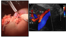
Figure 1. Dacron Graft Reconstruction of Right renal artery with postoperative renal Doppler ultrasound
To the best of our knowledge, there is no description of transplant renal artery reconstruction with a dacron graft in literature. There have been reports of two transplant RV and one RA reconstruction with Polytetrafluoroethylene (PTFE) graft with good outcomes [1,2]. A systematic meta-analysis of randomized controlled trials comparing Dacron® and PTFE as bypass materials for peripheral vascular surgery showed no evidence of an advantage of one over the other [3]. PTFE has been used for native renal artery reconstruction in patients with renal artery stenosis with good long-term results [4,5]. Most of these prosthetic vascular grafts exhibit reduced compliance compared to native arteries, which can potentially lead to dilatation, aneurysm, para-anastomotic pseudoaneurysm, and mechanical failure [6,7]. Takami, et al. showed that the knitted dacron graft diameter increases by 26%, compared with the package size, immediately after implantation and the graft further dilates 10.5%, compared with the diameter at discharge, and at 3.23% per year up to 5 years after surgery without any adverse events [8]. The long-term rate of dilatation in transplant settings where smaller grafts are used than in the studies mentioned above involving large diameter grafts to aorta, remains to be studied. With an expected
survival of over 12 years for living donor kidney transplants (https://optn.transplant.hrsa.gov/resources/guidance/kidney-donor-profile-index-kdpi-guide-for-clinicians/), theses grafts may actually outlive the kidney. Having these options available in OR can be extremely valuable in difficult cases, where no donor vein is readily available, and attempting to harvest other endogenous vessels like saphenous vein or internal iliac artery, could be time consuming, adding to cold ischemia time. However, long-term follow up is required to study the patency of prosthetic grafts in kidney transplantation.
Rough handling of donor renal artery during procurement or benching can lead to intimal injury with subsequent arterial dissection following implantation. In cases where no donor vessels are available, which is generally the case in living donors, dacron graft reconstruction can be achieved quickly and efficiently, with acceptable outcomes, since attempting to harvest an endogenous vessel could be time consuming adding cold ischemia time with the risk of delayed graft function and associated complications.
- Rough handling of donor renal artery during procurement or benching can lead to intimal injury with subsequent arterial dissection following implantation.
- In cases where no donor vessels are available, which is generally the case in living donors, dacron graft reconstruction can be achieved quickly and efficiently, with acceptable outcomes.
- Kandilis A, Di Cocco P, Rajagopa P, Hakim NS (2016) Salvage of a Live-Related Transplant Kidney Using an Interposition Polytetrafluoroethylene Vascular Graft: A Unique Case. Exp Clin Transplant 14: 679-681. [Crossref]
- Kamel MH, Thomas AA, Mohan P, Hickey DP (2007) Renal vessel reconstruction in kidney transplantation using a polytetrafluoroethylene (PTFE) vascular graft. Nephrol Dial Transplant 22: 1030-1032. [Crossref]
- Roll S, Müller-Nordhorn J, Keil T (2008) Dacron vs. PTFE as bypass materials in peripheral vascular surgery—systematic review and meta-analysis. BMC Surg 8: 22. [Crossref]
- Nghiem DD (2008) Use of spiral vein graft in living donor renal transplantation. Clin Transplant 22: 719-721. [Crossref]
- Paty PS, Darling RC 3rd, Lee D (2001) Is prosthetic renal artery reconstruction a durable procedure? An analysis of 489 bypass grafts. J Vasc Surg 34: 127-132. [Crossref]
- Tai NR, Salacinski HJ, Edwards A (2000) Compliance properties of conduits used in vascular reconstruction. Br J Surg 87: 1516-1524. [Crossref]
- Spadaccio C, Rainer A, Barbato R (2013) The fate of large-diameter Dacron® vascular grafts in surgical practice: are we really satisfied? Int J Cardiol 168: 5028-5029. [Crossref]
- Takami Y, Tajima K, Kato W (2012) Long-term size follow-up of knitted Dacron grafts (Gelseal™) used in the ascending aorta. Interact Cardiovasc Thorac Surg 14: 529-531. [Crossref]