Abstract
We present a case of papillary endothelial hyperplasia masquerading as a right ventricular mass in a patient with venous thromboembolism. This case highlights the known association of papillary endothelial hyperplasia with vascular stasis or thrombosis, and its rare occurrence in the right ventricle with associated saddle pulmonary embolus.
Keywords
Venous Thromboembolism, Electrocardiogram, Deep Vein Thrombosis, Pulmonary Embolism, Right Ventricle, Trans-thoracic Echocardiogram, Papillary Endothelial Hyperplasia
History of presentation
An 81-year-old woman with recent admission for ischemic stroke with residual left sided extremity weakness presented to the hospital with sudden onset of light-headedness and nausea with a 3-day history of right leg swelling and pain. She was in a routine outpatient physical therapy session when she developed positional light-headedness described as presyncope. She denied chest pain but endorsed dyspnea on exertion for a few days prior. She was administered intravenous normal saline by EMS en route which improved her light-headedness.
Upon presentation, she was afebrile, with a heart rate of 64, blood pressure of 150/70 and a respiratory rate of 24. She was hypoxic to 88% on room air at rest and with minimal ambulation, was profoundly hypoxic to 78% on room air. Oxygen supplementation was administered by nasal cannula. Examination revealed mild respiratory distress with tachypnea and normal jugular venous pulsations. Cardiac auscultation revealed normal first and second heart sounds without murmur or rub. Lung examination was clear to auscultation. Extremities were warm to touch with right leg pitting edema that was tender to palpation.
Past medical history
Hypertension, Diabetes Mellitus, Dyslipidemia, Ischemic stroke with residual left hemiparesis, Obstructive Sleep Apnea.
Differential diagnosis
The differential diagnosis at this point in the case although no confirmatory imaging, was still strongly favoring venous thromboembolism (VTE) as the etiology of presenting signs and symptoms.
The differential diagnosis of a right ventricular mass in the setting of VTE includes a thrombus in transit; primary or secondary cardiac tumors that could be benign or malignant in nature.
Investigations
Diagnostic work up in the emergency department revealed normal complete blood count and blood chemistry profile. High-sensitivity troponin was elevated at 829 ng/L. BNP was normal at 30 pg/ml. Electrocardiogram (EKG) revealed a normal sinus rhythm at rate 63, left ventricular hypertrophy, left axis deviation of the QRS complex, which is unchanged from her baseline EKG. Due to a high suspicion of an acute venous thromboembolic phenomenon, ultrasound venous duplex of bilateral lower extremities was performed that revealed a near-occlusive acute deep vein thrombosis (DVT) of the right popliteal, posterior tibial and peroneal veins. Urgent CT-angiogram of the chest revealed a large saddle pulmonary embolus (PE) beginning at the distal main pulmonary artery and extending into the right and left pulmonary arteries.Distal propagation of the thrombus on the right side into the 1st and 2nd order branches supplying the right upper lobe, right lower lobe, and right middle lobe was noted.Additional findings of a deviated interventricular septum towards the left, suggesting mild right heart strain were observed.A 1.9 cm low density mass within the right ventricle (RV) was noted which was suspicious of a thrombus in transit. An urgent transthoracic echocardiogram (TTE) was performed to further evaluate the RV mass which revealed a normal left ventricular ejection fraction of 60%, moderately dilated RV with a normal systolic function, moderate tricuspid regurgitation, and a 2.4 cm X 1.9 cm circular density in the RV apex that appeared as a filling defect with echo contrast administration (Figure 1). Abnormal flattening of the interventricular septum during systole was also noted that was consistent with RV pressure overload. Pulmonary artery systolic pressure was mildly elevated at 46 mm Hg. Bubble study during the TTE also revealed a right to left intra-cardiac shunt via the interatrial septum.
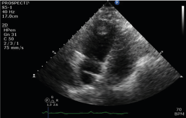
Figure 1.Four chamber TTE image showing right ventricular apical mass
Intra-operative resection of the RV mass revealed a 2.2 cm x 1.7 cm x 1.5 cm focally disrupted tan-pink soft tissue that weighed 2 grams (Figure 2). Subsequent pathological analysis of the tissue was consistent with papillary endothelial hyperplasia (PEH).
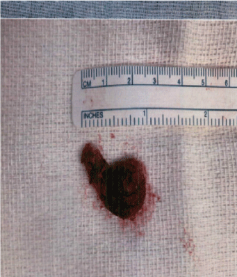
Figure 2.Right ventricular mass
Management
Supplemental oxygen therapy via nasal cannula was administered and the patient was admitted to the intensive care unit. Anticoagulation with intravenous heparin was initiated. Various treatment options of a saddle PE with hypoxia and evidence of right heart strain were considered such as systemic thrombolytic therapy, catheter directed thrombolysis or thrombectomy and surgical embolectomy. In order to address the RV mass that was concerning for a thrombus in transit in the setting of a high thrombus burden including DVT and a large saddle PE, the option of surgical embolectomy was chosen by a multidisciplinary team that included Cardiology, Intensivist and Cardiothoracic surgery services. Successful surgical embolectomy of the saddle PE measuring 32 cm x 1.2 cm was performed (Figure 3). Primary closure of the secundum atrial septal defect was also performed. Intraoperatively, the RV mass was found intertwined with the trabeculations and chordae of the tricuspid valve (Figure 4). It was 2.2 cm x 1.7cm x 1.5 cm in size and with a gross appearance of a myxoma, but the pathological evaluation was consistent with papillary endothelial hyperplasia (Figure 5). Patient recovered well and was discharged on apixaban for treatment of VTE.
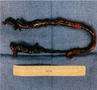
Figure 3.Resected saddle pulmonary embolus
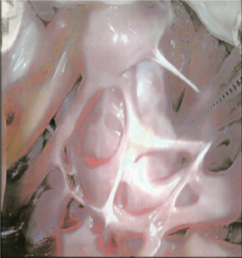
Figure 4.Intraoperative view of RV apex through the tricuspid valve.The mass is intertwined between trabeculae and is in close proximity to papillary muscle and chords
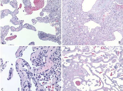
Figure 5.All 4 images of surgical pathology of RV mass displaying papillary formations with hyaline or fibrous stalks, anastomosing vascular channels, and plump endothelial cells.No necrosis, no atypia, no atypical mitotic figures are present
Discussion
PEH or Masson’s tumor is a rare, benign vascular lesion that can manifest as a soft tissue mass. It is believed to be due to a reactive proliferation of the endothelial cells forming papillary structures due to vascular trauma, stasis, or evolving thrombus[1]. It was first described in 1923 by Pierre Masson as hemangioendotheliomevegetantintravasculaire[2]. It is reported to occur in the vascular bed of skin, head, neck, and extremities. The diagnosis of PEH can be challenging as it requires a high index of suspicion and a thorough pathological evaluation of the tissue. Cardiac involvement appears to be extremely rare as per current literature review. Mantha etal. [3] reported a case of PEH in the left atrial appendage as a cause of cardioembolic stroke in a 70-year-old woman. Abad etal. [4] reported a case of cardiac hemangioma with findings of PEH in the left atrium. Safa etal. [5]reported a case of a one-month-old infant with hypoplastic left heart syndrome who was found to have coronary PEH causing a massive myocardial infarction.Abad et al.[6]reported a case of a left atrial hemangioma with PEH in a 42-year-old man.
This case of PEH involving the right ventricle has not been reported in the currently available published literature and it highlights the association of PEH and vascular thrombosis. The VTE phenomenon in our patient involving lower extremity DVT that embolized to the pulmonary artery traversing through the right ventricle perhaps served as a nidus for the formation of the Masson’s tumor in the RV. Surgical approach was opted in our patient mainly due to the high thrombus burden manifesting as a saddle PE and RV mass that was appropriately suspected to be a clot in transit given the clinical context.
Follow up
Patient continued to do well post discharge as evaluated several times in the outpatient setting. Cardiac Magnetic Resonance Imaging was performed which did not reveal any residual or recurrence of an intra-cardiac mass. There was no delayed enhancement in the myocardium to suggest occult scarring, infarction, inflammatory or infiltrative disease. A repeat TTE performed 3 months after the index presentation did not reveal an intra-cardiac mass.
Conclusion
PEH or Masson’s tumor can very rarely present as an intra-cardiac mass. Vascular stasis or thrombosis can predispose to reactive endothelial hyperplasia. In the setting of a VTE phenomenon, a right sided cardiac mass can be misdiagnosed as a thrombus in transit. Surgical exploration and resection may be needed for the evaluation of gross morphology and confirmation of the pathological diagnosis This case highlights the association between venous thromboembolism and right ventricular PEH.
Learning objective
Intra-cardiac PEH is a very rare differential diagnosis of an RV mass. A high index of suspicion in the appropriate clinical context with vascular stasis or thrombosis is warranted to pursue surgical diagnosis and excision. Data regarding the sequelae of undiagnosed or untreated intra-cardiac PEH is lacking in the current literature.
Funding support and author disclosure
The authors have reported that they have no relationships relevant to the contents of this paper to disclose.
References
- Mahapatra, Qury S (2015) Intravascular papillary endothelial hyperplasia: An unusual histopathological entity. Indian Dermatol J 6: 277-279. [Crossref]
- Hong, Seok-Gi (2004) Intravascular papillary endothelial hyperplasia (Masson's hemangioma) of the liver: a new hepatic lesion. J Korean Med Sci 19: 305-308. [Crossref]
- Mantha Y, Harada R, Hieda M (2020) Masson Tumor in the Left Atrial Appendage Presenting as Cardioembolic Cerebral Infarction. J Am Coll Cardiol Case Rep2: 1969-1973.[Crossref]
- Abad C, Campo E, Estruch R, Condom E, Barriuso C, et al. (1990) Cardiac hemangioma with papillary endothelial hyperplasia: report of a resected case and review of the literature. Ann ThoracSurg 49:305-308. [Crossref]
- Safa R, Garcia R,Delius R,Kaur G, Youssef L, et al.(2019) Intravascular Papillary Endothelial Hyperplasia in the Coronary Artery: An Unusual Cause of Massive Myocardial Infarction in Hypoplastic Left Heart Syndrome. FetalPediatrPathol 38: 511-517.[Crossref]
- Abad C, Campo E, Estruch R, Condom E, Barriuso C, et al. (1990) Cardiac Hemangioma With Papillary Endothelial Hyperplasia: Report of a Resected Case and Review of the Literature, Department of Cardiovascular Surgery, Pathology, and Medicine, Hospital CLinico, Barcelona, Spain. Ann ThoracSurg49:305-308. [Crossref]