Abstract
Rosai-Dorfman is a rare disease, benign, proliferative lesion that frequently affects young adults, characterized by the presence of large histiocytes with emperipolesis. The most common presentation is painless cervical lymphadenopathy, with extranodal involvement being uncommon. In the breast the disease is quite rare, with few reports in English literature. We reported a unique case of a 46-year-old patient with a left breast nodule with imaging tests mimicking breast cancer. She was submitted to a left breast section for excision of the nodule, sending material for the pathology and immunohistochemistry in which she confirmed Rosai-Dorfman's disease. The patient was followed up for 48 months, without complaints, with imaging exams without evidence of disease recurrence after surgery.
Key words
breast, histiocytosis sinus, Rosai Dorfman, non-Langerhans cell histiocytosis
Introduction
Rosai-Dorfman disease is a rare, idiopathic, benign, proliferative lesion that affects histiocytes, recognized as a distinct entity since 1969 when it was reported by Rosai and Dorfman. Its histopathological features are similar regardless of where they occur. The most common presentation is painless cervical lymphadenopathy, but extranodal involvement is not uncommon, reported in up to 43% of cases [1]. It has been reported that 23% of cases have exclusively extranodal involvement. In the breast the disease remains a rare entity, with about 27 cases reported in the English literature. Because Rosai-Dorfman's disease often mimics invasive breast carcinoma in its clinical presentation and radiographic appearance - and can mimic other histiocytic lesions microscopically - awareness and appropriate diagnosis of this entity are essential for proper treatment.
Methodology
This is a case report through a retrospective analysis of the patient's data collection and clinical, pathological and image examinations obtained in the Liga Norteriograndese Contra Câncer - Natal-RN (Brazil) of the patient under study. The review period is from November 2013 to November 2017.
Case
J.C.S, female, 46 years old, sought the mastology service of the Liga Norteriograndense Contra Cancer - Brazil report on the appearance of a nodule in the left breast in August 2013. Nulliparous patient, without other known risk factors for breast cancer and without history family relationship. At the physical examination, mobile and regular nodulation of 4cm was palpated in the lateral superior quadrant (LSQ) of the left breast (LB), axillae without palpable nodulations. Mammography showed a dense nodule of 5.0×4.0 cm in QSL of ME, classified as Bi-rads 0. Ultrasonography revealed a heterogeneous area of poorly defined contours with focal architectural alteration, 3.3×2.9cm 1h to 10.5cm of the left nipple, Bi-rads 4. It was submitted to fine needle aspiration (FNA), whose cytology showed absence of ductal cells and many mature lymphocytes and some immature lymphocytes. Patient was submitted to sectionectomy of ME for palpable nodule excision. Anatomopathological and immunohistochemical study evidenced Rosai-Dorfman's disease. Patient has been in clinical follow-up for 48 months, with no evidence of recurrence of breast disease or elsewhere (Figure 1).
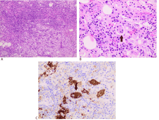
Figure 1 (A-C). Infiltrate constituted by plasmocytes, lymphocytes and histiocytes with ample cytoplasm, vesiculous nuclei and with presence of lymphocytes and intact plasmocytes in the cytoplasm (emperipolese)
Discussion
Rosai-Dorfman disease (DRD), also known as sinus histiocytosis or non-Langerhans cell histiocytosis, is an idiopathic disorder that often affects children or young adults, although it can affect any age group [2] and is characterized by the presence of large histiocytes with emperipolesis [3]. Nearly 90% of the patients with DRD present involvement of cervical lymph nodes, although all the organs can be involved.
The etiology and pathogenesis of this rare disease are not well known. A cytokine-mediated migration of monocytes may be involved in the accumulation and activation of histiocytes. This functional activation could be triggered by different stimuli, due to the coexistence of DRD and autoimmune diseases, haematological neoplasias and postinfectious conditions. Viruses such as Herpesvirus 6 and Epstein-Barr virus were implicated as potential causative agents, however, without strong evidence [4].
The classic presentation for Rosai-Dorfman disease is bilateral painless cervical lymphadenopathy in an asymptomatic young patient, or with symptoms such as fever and weight loss [4]. The extranodal involvement due to DRD was documented in 43% of the cases [1], with the most frequent sites being skin and soft tissues, upper respiratory tract, oral cavity, genitourinary tract and bone [5]. Involvement of the head and neck was reported in 22% of cases, most commonly the nasal cavity, followed by the parotid gland [4]. Laboratory tests may show a high erythrocyte sedimentation rate in most patients. Less commonly, there may be anaemia, leucocytosis and polyclonal hypergammaglobulinemia [6].
DRD limited to the breast is extremely rare; the patients typically have palpable painful masses in the breast, but many cases of asymptomatic women are only discovered on imaging tests. They usually manifest as ill-defined or irregular masses without mammography calcifications, or hypoechoic masses on ultrasound, but may have an indistinguishable appearance of breast carcinoma in these examinations.
Hormically, DRD presents as a dense lymph histiocytic infiltrate containing layers of histiocytes with small nuclei and foamy cytoplasm. Emperipolesis (intact lymphocytes within histiocytes) is present but not easily seen in extranodal disease. In immunohistochemistry, histiocytes show reactivity for S-100 and CD68 proteins, in addition to CD163, á1-antiquimotrypsin, á1-antitrypsin, fascine and HAM-56, whereas CD1a is typically negative. Lymph nodes, if involved, show dilation of nodal sinuses filled by histiocytes, lymphocytes and plasma cells, and pericapsular fibrosis. DRD lesions present moderate expression of IL-6, which may be related to the presence of associated polyclonal plasmacytosis and hypergammaglobulinemia. In addition, the lesions tend to strongly express IL-1â and TNF-á. Systemic symptoms may be related to the increased production of these cytokines [4,7].
The disease must be histologically differentiated from Langerhans cell histiocytosis (HCL), infectious and lymphoproliferative diseases and sinus hyperplasia. Positivity for S-100 can generally distinguish between the latter condition and DRD.
Differential diagnosis based on physical exam characteristics includes diseases that cause benign and malignant mammary nodules. Regarding the histopathological characteristics, DRD should be differentiated from carcinoma, diabetic mastopathy, fatty necrosis, granulomatous mastitis and Langerhans histiocytosis. The conclusive diagnosis may be suggested by fine needle aspiration and established by thick needle biopsy or surgical excision.
The clinical course of DRD is unpredictable, with episodes of exacerbation and remissions. The disease is often self-limited and of good prognosis, but about 5-11% of patients die from DRD [4]. There are 3 distinct evolutions of the disease: 1) sudden lymphadenomegaly, with spontaneous regression and without other recurrences; 2) patients with immunological abnormalities at presentation presenting more disseminated nodal disease and higher mortality; 3) patients with multinodal and extranodal disease, with prolonged clinical course with multiple relapses and remissions for years. In these cases, the severity of the disease depends on the type and number of extranodal sites. The pattern of breast DRD is uncertain, due to the limited number of cases studied in the literature [4,7].
In most cases, DRD has a benign course and treatment is not necessary and is indicated for patients with extranodal disease involving vital organs or those with nodal disease causing potentially fatal complications. Surgery is usually limited to biopsy, but cytoreductive surgery may be necessary in patients with vital organ involvement when feasible, or partial resection with radiosurgery and / or radiotherapy in larger lesions, with limited results. Patients with systemic or sudden-onset lymph node symptoms may be treated with prolonged low-dose corticosteroid treatment. The use of chemotherapy and immunosuppressive drugs should be restricted to patients with potentially fatal illness or in nonresponsive or relapsing cases, but further studies are needed to determine the best treatment strategy [4,8].
The surgical management of mammary DRE should be based on the need for differential diagnosis of neoplastic lesions. The follow-up of these patients is aimed at observing remission of the disease or relapse and finding other sites of extramammary disease, although there is no current consensus in the literature about such practice.
In this clinical case,2021 Copyright OAT. All rights reserv DRD sites and is in a 48-month clinical follow-up free of disease, conferring an excellent prognosis without any additional treatment.
Conclusion
Rosai-Dorfman disease of the breast is commonly confused with a malignant neoplasm of the breast in the initial approach, both in clinical and radiological presentation and pathologists should be familiar with the histopathological features of this entity. Thus, knowledge of this disease and adequate diagnosis are essential for the appropriate treatment. The most commonly used approach is surgical excision, with variable rates of recurrence. Their course is generally favourable. Further studies are expected to define the best therapeutic strategy for such patients.
References
- Pham CB, Abruzzo LV, Cook E, Whitman GJ, Stephens TW (2005) Rosai-Dorfman disease of the breast. AJR Am J Roentgenol 185: 971-972.
- Juskevicius J, Finlay JL (2001) Rosai-Dorfman disease of the parotid gland, cytologic and histopathologic findings with immunohistochemical correlation. Arch Pathol Lab Med 125: 1348-1350. [Crossref]
- Cohen AF, Haroche J, Emile JF, Charlotte F, Barete S, et al. (2018) Rosai-Dorfman disease: Diagnosis and therapeutic challenges. Rev Med Interne. [Crossref]
- Histiocyte Society (2011) Rosai-Dorfman Disease Review.
- Foucar E, Rosai J, Dorfman R (1990) Sinus histiocytosis with massive lymphadenopathy (Rosai-Dorfman disease): review of the entity. Semin Diagn Pathol 7: 19–73. [Crossref]
- Hummel P, Waisman J, Chhieng D, Yan Z, Cohen JM, et al. (1999) Fine-needle aspiration cytology of Rosai-Dorfman disease of the breast: a case re- port. Diagn Cytopathol 21: 287–291. [Crossref]
- Warnke RA, Weiss LM, Chan JKC, Cleary ML, Dorfman RF, et al. (1995) Tumors of the lymph nodes and spleen. In: Armed Forces Insti- tute of Pathology, Atlas of tumor pathology, (3rd ed.), fasc. 14. Washington, DC: AFIP, pp: 349–360.
- Morkowski JJ, Nguyen CV, Lin P, Farr M, Abraham SC, et al. (2010) Rosai-Dorfman disease confined to the breast. Ann Diagn Pathol 14: 81-87. [Crossref]