A 63 years old man with history of diabetes and hypertension, presented for bone pain. Pelvic X Ray showed multiple osteoconsensant lesions of the iliac wings, the sacrum and the femurs (Figure 1). CT scan objectified multiple hepatic metastases with a laterorectal mass (Figure 2). Colonoscopy showed aspect of extrinsic compression. Endoscopic ultrasound objectified a rectal mucosal lesion of 5x3,5 centimeters appearing in contact with the prostate (Figure 3) [1-5]. Histologically, hepatic biopsy confirmed a tumor proliferation that was made of two architectural aspects independent cells, and trabeculolobular classical endocrine architecture with expression of synaptophysin on immunochemistry [1-5] (Figure 4).
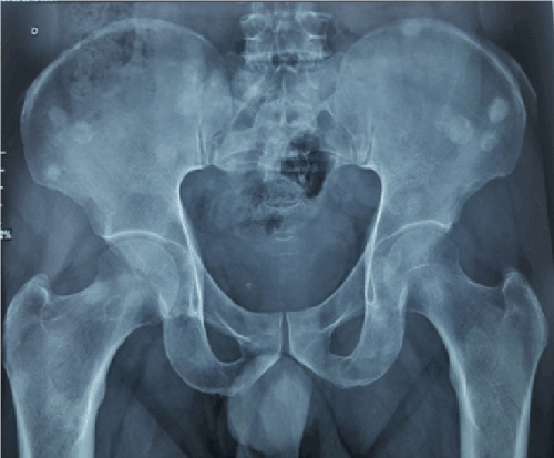
Figure 1. Pelvic X Ray showed multiple osteoconsensant lesions of the iliac wings, the sacrum and the femurs
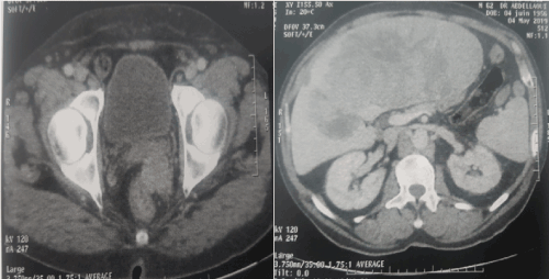
Figure 2. CT scan objectified multiple hepatic metastases with a laterorectal mass
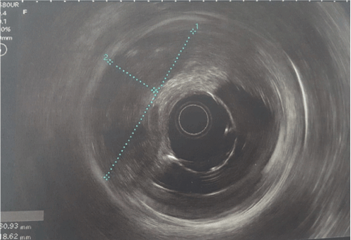
Figure 3. Endoscopic ultrasound objectified a rectal mucosal lesion of 5x3,5 centimeters appearing in contact with the prostate
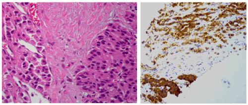
Figure 4. hepatic biopsy showing a tumor proliferation that was made of two architectural aspects independent cells, and trabeculolobular classical endocrine architecture with expression of synaptophysin on immunochemistry
- Haq S, Batra VV, Majumdar K, Javed A, Agarwal AK, et al. (2015) Signet ring cell neuroendocrine tumor liver with mesenteric metastasis: Description of a rare phenomenon, with literature review. J Cancer Res Ther 11: 658. [Crossref]
- Madakshira MG, Radotra BD, Singh V (2019) Signet ring: A rare morphology of metastatic neuroendocrine tumor. Indian J Pathol Microbiol 62: 335-336. [Crossref]
- Shaieb W, Krishna K, Kim S, Godman M, Rock J, et al. (2016) Appendiceal Neuroendocrine, Goblet and Signet-Ring Cell Tumors: A Spectrum of Diseases with Different Patterns of Presentation and Outcome. Cancer Res Treat 48: 596-604. [Crossref]
- He F, Nie L (2019) Synchronous neuroendocrine tumor and signet-ring cell carcinoma in the stomach. Clin Res Hepatol Gastroenterol 43: 505-7.
- Korphaisarn K, Morris V, Davis JS, Overman MJ, Fogelman DR, et al. (2019) Signet ring cell colorectal cancer: genomic insights into a rare subpopulation of colorectal adenocarcinoma.Br J Cancer 121: 505-10. [Crossref]
Editorial Information
Editor-in-Chief
Yi-Hwa Liu
Yale University
Article Type
Case Report
Publication history
Received date: October 02, 2019
Accepted date: October 09, 2019
Published date: October 11, 2019
Copyright
©2019 Sabbah M. This is an open-access article distributed under the terms of the Creative Commons Attribution License, which permits unrestricted use, distribution, and reproduction in any medium, provided the original author and source are credited.
Citation
Sabbah M, Trad D, Bellil N, Jouini R, Ouakaa A, et al. (2019) Signet ring cell neuroendocrine tumor. Radiol Diagn Imaging 3: DOI: 10.15761/RDI.1000161
Corresponding author
Sabbah Meriam
Habib Thameur Hospital, Rue El Messelekh, Montfleury, Tunis, Tunisia
E-mail : bhuvaneswari.bibleraaj@uhsm.nhs.uk
Figure 1. Pelvic X Ray showed multiple osteoconsensant lesions of the iliac wings, the sacrum and the femurs
Figure 2. CT scan objectified multiple hepatic metastases with a laterorectal mass
Figure 3. Endoscopic ultrasound objectified a rectal mucosal lesion of 5x3,5 centimeters appearing in contact with the prostate
Figure 4. hepatic biopsy showing a tumor proliferation that was made of two architectural aspects independent cells, and trabeculolobular classical endocrine architecture with expression of synaptophysin on immunochemistry