Abstract
The dental, craniofacial and systemic anomalies in a case with the solitary maxillary central incisor (SMCI) and short stature syndrome are described. The patient, a 9.3- year-old female, manifested SMCI and congenital absence of the permanent maxillary lateral and the four mandibular incisors. Main craniofacial anomalies were microcephaly, brachycephaly, platybasia, underdeveloped anterior and posterior cranial base and maxilla, hypotelorism, short and narrow upper face, and low set and prominent ears. On cephalometrics the reported syndrome was found to be quite distinct from other syndromes. Systemic manifestations included bilateral hip dislocation, double kidney, and short stature with normal
growth hormone values. The lack of information for genetic counseling of individuals with the SMCI and short stature syndrome is discussed.
Key words
multiple abnormalities, hypodontia, short stature, cephalometrics
Introduction
Solitary maxillary central incisor has been described as an isolated abnormality in idividuals with normal stature and growth hormone levels [1,2], in association with short stature and isolated growth hormone deficiency, "monosuperocentroincisivodontic dwarfism" [3-5], with short stature but no growth hormone abnormalities [4,6,7], and in other reports, where growth hormone levels were not presented [8-13]. Solitary maxillary central incisor has also been described in association with del(18p) syndrome [14,15], with precocious puberty and hypothalamic hamartoma [16] , with short stature, excessive ADH secretion, and hyponatremia [17], in hypomelanosis of Ito with iris coloboma [18] and with unusual forms of ectodermal dysplasia [19,20]. It may occur on occasion as a striking microform of holoprosencephaly, most commonly in the autosomal dominant (A.D) form [21-28], and along with absent maxillary incisors are very common in holoprosencephaly with severe facial
dysmorphism [29,30].
A case with the syndrome of solitary maxillary central incisor and short stature and cephalometric analysis of the patient is presented.
Case report
The patient, a 9.3 year-old girl, was delivered by cesarean section, because of breech presentation, after a
full-term, uncomplicated pregnancy, with no exposure to radiation or any unusual agent. Birth length (51 cm), weight (3.600 gr) and head circumference (33 cm) were normal. The APGAR score was 6. As a neonate she was hospitalized in an infant incubator for 2 days for respiratory distress. Her growth and developmental milestones were retarded. At 3.5 months she was hospitalized for failure to grow and gain weight. She had bilateral hip dislocation and double kidney on the right side, for which she had heminefrectomy at 18 months. CT brain scan at 18 months disclosed no brain dysmorphology. Measurments on a P-A cephalogram taken at the age of 4 10/12 showed severe hypotelorism (bony interorbital distance 12 mm, approximately -5 S.D.) and wide neurocranium (cranial breadth 17.5 cm, +3.5 S.D.). Smell was reported to be functional. Peak growth values post Arginine and glucagon were 5.78 and 12 ng/ml, respectively (normal>10). Delayed eruption of a comlete, but with single maxillary central incisor, primary dentition was reported. No consanguinity and similar case in the family was
mentioned. The target height was 154+4.5 cm.
On physical examination at age 9.3 years her height was 117 cm (SDs -3.1), and her weight 21 Kg (< 5th
centile). She had microcephaly (head circ.: 50 cm), brachycephaly (ceph. index: 83.8), groove along the
coronal suture, low set and prominent ears, hypotelorism, short and narrow upper face, hypoplastic maxilla, narrow, mixed dentition with single maxillary permanent central incisor (Figure 1) highly arched palate (Figure 2), and hyperextensibility of joints. She demonstrated a borderline mentality. Measurments on casts (age 10.6) showed a short premaxilla (1.1 cm), narrow palate (palatal width 2.5 cm, -2,5 S.D.), high palate (palatal height 1.75 cm, >2S.D.??). A panoramic radiograph (done at age 8) showed hypodontia of the permanent dentition; the maxillary lateral and the four mandibular incisors were congenitally issing (Figure 3).
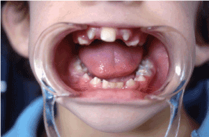
Figure 1. Patient's dentition on admission. Single maxillary central incisor, absent permanent lateral upper
incisors, retention of primary mandibular incisors
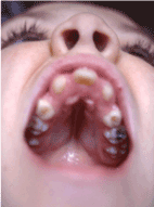
Figure 2. Patient's palate, highly arched
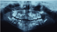
Figure 3. Panoramic radiograph of the patient at the age of 8. Absence of the permanent maxillary lateral incisors and the four mandibular incisors
Cephalometric analysis (Table 1) revealed severely decreased anterior and posterior cranial base, severely
increased cranial base saddle angle (platybasia), severely tilted up anterior cranial base, severely short anterior and posterior upper facial height, severely short maxilla, severely retruded maxilla, concave profile, severely retruded maxillary denture base, well positioned mandible relatively to the forehead, and
mildly retruded mandibular denture base (Figure 4).
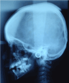
Figure 4. Lateral cephalometric radiograph of the patient. Platybasia, concave profile (see text)
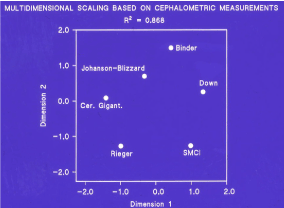
Figure 5. SMCI and short stature syndrome quite distinct from the other five syndromes
Chomosomal analysis,including G-banding, was normal (46,XX).
Discussion
We described the clinical and cephalometric phenotypic features of a female patient with the syndrome of
solitary maxillary central incisor and short stature, with normal growth hormone levels. The absence of the
permanent maxillary lateral incisors and, more strikingly, all four mandibular incisors denotes that the developmental defect in the median structures of the face is more severe in our case than in the reported so far in the literature, since none of those had this hypodontia in addition to the single maxillary central incisor. The girl also had microcephaly, severe hypotelorism (interpapillary distance 44 mm, sufficiently below the 3rd percentile), and borderline mentality. Although smell was reported to be functional and a CT brain scan at the age of 18 months disclosed no brain dysmorfology, the essential question of a minor defect of the midline structures of the forebrain not detected by the scanning still remains. It has been emphasized that SMCI may occur alone or in other conditions bearing no relationship to holoprocencephaly. Nevertheless, the pathogenetic similarity of the SMCI and short stature syndrome to holoprosencephalic microforms is obvious, although no etiologic relationship has been established. However, individuals with SMCI with or without hypotelorism, or microcephaly in families with A.D. form of holoprosencephaly had children with holoprocencephaly. On the other hand, all the reported cases with the syndrome of SMCI and short stature were sporadic, all but one female who didn't have any pregnancies, had not reached reproductive age and there is no follow-up information about their reproduction. Nothing is known about the genetics and the recurrence risk of the syndrome. So the risk for individuals with the syndrome of having holoprosencephalic children is not known.
In order to give answers to these questions and more information on the phenotype of the syndrome we did cephalometric analysis of the patient, not done to any of the previously reported cases. In addition, by using the "multidimensional scaling" statistical technique, we tried to examine differences or similarities among syndromes for all characheristic cephalometric measurements found in our case, collecting data from the literature concerning such measurments in other syndromes. Unfortunatelly, there were not enough data for all the other syndromes in all the measurements to be included in this analysis, including five valuable cases of DeMyer (holoprosencephaly) sequence 31 , which could be very informative. Finally, only six measurments: 1) anterior and 2) posterior cranial base, 3) cranial base- saddle angle, 4) maxillary length, 5) maxillary protrusion and 6) mandible protrusion, of six syndromes namely: SMCI and short stature (our case), Rieger 32 , Binder 33, 34 , Johanson-Blizzard 35 , cerebral gigantism
36 and Down 37 were used in the analysis.
The results were clear cut. First, the model fit very well the data, that is the multidimensional scaling captured successfully the differences among syndromes in the cephalometric space; this is shown by the high coefficient of determination, R2=0.868. Second, the 2- dimensional plot (Figure 5) shows clearly that the syndrome of SMCI and short stature is quite distinct from the other five syndromes. In fact, it is more so than some other syndromes are distinct among themselves. This shows that cephalometric measurements are indeed a powerful tool in distinguishing syndromes.
Conclusion
For the future, we need more cases of the syndrome of SMCI and short stature, and cases of the A.D. form of holoprosencephaly covering the wide spectrum between the two extreme manifestations of the gene expression, i.e, holoprosencephaly and SMCI, to be cephalometrically analysed. That will, at least partly, elucidate the pathogenesis of the SMCI and short stature syndrome, whether it is simply a craniofacial dysplasia or a cerebral craniofacial dysplasia and give us information for the genetic counseling.
Acknowledgements
We wish to acknowledge the contribution of Dr. Apostolos Georgopoulos for statistical advice concrning the multidimensional analysis and to thank Dr. Robert Gorlin for reviewing the primary form of the paper.
References
- Wesley RK, Hoffman WH, Perrin J, Delaney JR Jr (1978) Solitary maxillary central incisor and normal stature. Oral Surg Oral Med Oral Pathol 46: 837-842. [Crossref]
- Santoro FP, Wesley RK (1983) Clinical evaluation of two patients with a single maxillary central incisor. ASDC J Dent Child 50: 379-381. [Crossref]
- Rappaport EB, Ulstrom R, Gorlin RJ (1976) Monosuperocentroincisivodontic dwarfism. Birth Defects Orig Artic Ser 12: 243-245. [Crossref]
- Rappaport EB, Ulstrom RA, Gorlin RJ, Lucky AW, Colle E, et al. (1977) Solitary maxillary central incisor and short stature. J Pediatr 91: 924-928. [Crossref]
- Vanelli M, Bernasconi S, Balestrazzi P (1980) [Solitary maxillary central incisor with growth hormone deficiency (author's transl)]. Arch Fr Pediatr 37: 321-322. [Crossref]
- Hayward JR (1979) Observations on midline deformity and the solitary maxillary central incisor syndrome. J Hosp Dent Pract 13: 113-114. [Crossref]
- Parker PR, Vann WF Jr (1985) Solitary maxillary central incisor: clinical report. Pediatr Dent 7: 134-136. [Crossref]
- Scott DC (1958) Absence of upper central incisor. Br Dent J 104: 247-248.
- Fulstow ED (1968) The congenital absence of an upper central incisor. Report of a case. Br Dent J 124: 186-188. [Crossref]
- Kopp WK (1967) A hereditary congenitally missing maxillary central incisor. Oral Surg Oral Med Oral Pathol 24: 367.
- Ellisdon PS, Marshall KF (1970) Connation of maxillary incisors. Br Dent J 129: 16-21. [Crossref]
- Holm AK, Lundberg L (1972) Hypodontia of both primary and permanent central upper incisor. Description of a case. Odontol Revy 23: 429-436. [Crossref]
- Bazan MT (1983) Fusion of maxillary incisors across the midline: clinical report. Pediatr Dent 5: 220-
221.
- Dolan LM, Willson K, Wilson WG (1981) 18p - syndrome with a single central maxillary incisor. J Med Genet 18: 396-398. [Crossref]
- Aughton DJ, AlSaadi AA, Transue DJ (1991) Single maxillary central incisor in a girl with del(18p) syndrome. J Med Genet 28: 530-532. [Crossref]
- Winter WE, Rosenbloom AL, Maclaren NK, Mickle PJ (1982) Solitary central maxillary incisor associated with precocious puberty and hypothalamic hamartoma. J Pediatr 101: 965-967. [Crossref]
- Friedman AL, Chesney RW, Bargman GJ, Segar WE (1979) Lack of inhibition of vasopressin release in midfacial hypoplasia. J Pediatr 94: 591-594. [Crossref]
- Bartholomew DW, Jabs EW, Levin LS, Ribovich R (1987) Single maxillary central incisor and coloboma in hypomelanosis of Ito. Clin Genet 31: 370-373. [Crossref]
- Winter RM, MacDermot KD, Hill FJ (1988) Sparse hair, short stature, hypoplastic thumbs, single upper central incisor and abnormal skin pigmentation: a possible "new" form of ectodermal dysplasia. Am J Med Genet 29: 209-216. [Crossref]
- Buntinx I, Baraitser M (1989) A single maxillary incisor as a manifestation of an ectodermal dysplasia. J Med Genet 26: 648-651. [Crossref]
- Patel H, Dolman CL, Byrne MA (1972) Holoprosencephaly with median cleft lip. Clinical, pathological, and echoencephalographic study. Am J Dis Child 124: 217-221. [Crossref]
- Cohen MM Jr (1974) Letter: Holoprosencephaly revisited. Am J Dis Child 127: 597. [Crossref]
- Lowry RB (1974) Letter: Holoprosencephaly. Am J Dis Child 128: 887. [Crossref]
- Berry SA, Pierpont ME, Gorlin RJ (1984) Single central incisor in familial holoprosencephaly. J Pediatr 104: 877-880. [Crossref]
- Hattori H, Okuno T, Momoi T, Kataoka K, Mikawa H, et al. (1987) Single central maxillary incisor and holoprosencephaly. Am J Med Genet 28: 483-487. [Crossref]
- Fryns JP, Van den Berghe H (1988) Single central maxillary incisor and holoprosencephaly. Am J Med Genet 30: 943-944. [Crossref]
- Ardinger HH, Bartley JA (1988) Microcephaly in familial holoprosencephaly. J Craniofac Genet Dev Biol 8: 53-61. [Crossref]
- Jaramillo C, Brandt SK, Jorgenson RJ (1988) Autosomal dominant inheritance of the DeMyer Sequence. J Craniofac Genet Dev Biol 8: 199-204. [Crossref]
- Cohen MM Jr (1989) Perspectives on holoprosencephaly: Part III. Spectra, distinctions, continuities, and discontinuities. Am J Med Genet 34: 271-288. [Crossref]
- Gorlin RJ, Cohen MM Jr, Levin LS (1990) Syndromes of the head and neck. New Yrk, Oxford: Oxford University Press: 873.
- Spolyar JL, Eldis F, Benjamins D (1991) Five cases of DeMyer sequence: An interophthalmic dysplasia. Cleft Palate- Craniofac J 28: 103-113. [Crossref]
- Brooks JK, Coccaro PJ, Zarbin MA (1989) The Rieger anomaly concomitant with multiple dental, craniofacial, and somatic midline anomalies and short stature. Oral Surg Oral Med Oral Pathol 68: 717-724. [Crossref]
- Olow-Nordenram M, Thilander B (1987) The craniofacial morphology in individuals with maxillonasal dysplasia (Binder's syndrome). A longitudinal cephalometric study of orthodontically treated children. Eur J Orthodont 9: 224-236.
- Horswell BB, Holmes AD, Levant BA, Barnett JS (1988) Cephalometric and anthropomorphic observations of Binder's syndrome: a study of 19 patients. Plast Reconstr Surg 81: 325-335. [Crossref]
- Motohashi N, Pruzansky S, Day D (1981) Roentgencephalometric analysis of craniofacial growth in the Johanson- Blizzard syndrome. J Craniofac Genet Dev Biol 1:57-72.
- Motohashi N, Pruzansky S, Kawata T (1981) Roentgencephalometric analysis of cerebral gigantism: report of four patients. J Craniofac Genet Dev Biol 1: 73-94. [Crossref]
- Fischer-Brandies H (1988) Cephalometric comparison between children with and without Down's syndrome. Eur J Orthod 10: 255-263. [Crossref]
- Riolo ML, Moyers RE, McNamara JA, Hunter WS (1974) An atlas of craniofacial growth: cephalometric standards from the University school growth study. Ann Arbor: University of Michigan. (Craniofacial growth series, Center for Human Growth and Development, monograph 2).