Abstract
The congénial dermal sinus is usually considered as marker of underlying sinal dysraphism and tuft of hairs are also commonly present. However, the cord may split into two hemicords, contained in a single dural or seperate dural sheath, may be associated with fibrous or bony spur causing tethering and may have associated thickened or fatty filum terminale. Authors report a first cases in the western literature, where one split hemicord had demoid cysto and other cord was pushed laterally and had thickened fatty filum terminale. She was managed with surgery sucessfully. Clinical feature, neuroimaging and surgical managent along with pertinent literature is reviewed brieflly.
Key words
hemicord, dermoid cyst of one hemicord, dermal sinus, tethered cord
Introduction
The congenital dermal sinus is a type of closed spinal dysraphism in which an epithelium-lined sinus tract from the dorsal skin surface extends inwards for a variable distance. It occurs during neurulation when the neural groove closes to form the neural tube on 26 days of gestation and results from a failure of neuroectoderm to separate from the cutaneous ectoderm.
Dermal sinus is a congenital lesion found most frequently in lumbosacral region followed by occipital region. Its association with intramedullary lesion is very rare. Author report a case of spinal dysraphism with dermal sinus associated with intramedullary dermoid sinus.
Case report
An 11- year -old school girl delivered at term, to a non-consaguinous parents. Parent noticed presence of dermal sinus with tufts of hair with occasional watery discharge since birth in lumbosacral area .For last one year prior to current admission in hospital, she also noted complaint of low back ache. She had good bladder and bowel control. On admission, vitals were stable, neurological evaluation showed no motor or sensory deficit. Local examination showed presence of a dermal sinus containing clear watery discharge along with tuft of hairs in lumbosacral region. Lab investigations revealed haemoglobin was 10.8 g/dl, leucocyte count 13,900 with platelets count -1,13,000 and rest of heamatological and serum biochemistry were within normal limit. MRI of lumbosacral spine showed presence of two hemicord (Figure 1 and 2) with dermoid cyst at L2-3 vertebral level with fatty filum terminate (Figure 3).
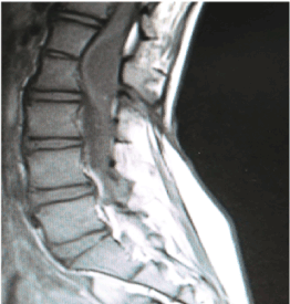
Figure 1. Sagital section, t1 weighted magnetic resonanace imaging showing hypointense dermiod cyst
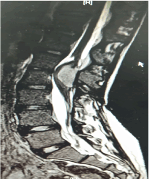
Figure 2. Sagital section, t2 weighted magnetic resonanace imaging showing hypointense dermiod cyst
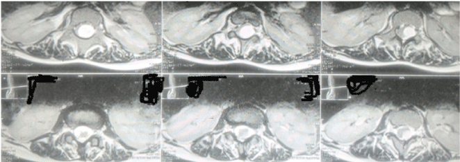
Figure 3. Axial section, t2 weighted magneic resonanace imaging showing hypointense dermiod cyst
She underwent surgery under general anaesthesia in the prone position. A vertical midline skin incision encircling the skin dimple was made, and the sinus tract was followed to the spinous process of L2 with the dissection of the dermal sinus stalk. The sinus tract traversed the lamina of L2 and then pierced the duramater. L1-L3 laminoplasty was performed, and the dura was opened. Two hemicords were found and a well-encapsulated, cystic, intramedullary tumor containing wax-like sebaceous materials was encountered in communication with the sinus tract in one hemicord while other hemicord was lying separately, having thickened filum. With microsurgical technique, total surgical microscope made it possible to find a plane between the tumor and the spinal cord, and total excision was performed. Detethering was done in another hemicord in which the filum was thickened. The dura was closed in a water-tight fashion and wound was closed in layers. Postoperatively patient was neurologically intact. There were no post op complications like CSF leak, pseudomeningocoele etc. She was discharged on fourth day following surgery. At last follow -up after two years after surgical procedure, she was doing well.
Discussion
Walter and Bucy first described congenital dermal sinus in 1932 [1]. It is basically formed due to defective separation of cutaneous ectoderm with neuroectodermal during the event of neurulation. Most frequently found in lumbosacral area followed by occipital area. Dermal sinus is considered as a marker of spinal dysraphism.
Dermoids are rare, benign, slow-growing lesions constituting 1.1% of intraspinal tumors [2]. The majority are in the extramedullary or subdural juxtamedullary location. Intraspinal dermoid occurs in the lumbosacral region, usually in the conus or cauda equina. Wang et al. described a case of congenital dermal sinus with T6-T7 level dermoid [3].
Magnetic resonance imaging provides rapid and accurate identification of the extent of lesions, clearly delineates the extraspinal portion of the sinus tract and clearly demonstrates presence of associated inclusion tumor, and defines the degree of the spinal cord compression, presence of fatty filum terminale, presence of spur and its nature and helps in surgical planning. The typical characteristics of dermoid cyst on magnetic resonance imaging include hypointense signal on T1-weighted image, hyperintense on T2-weighted image, with peripheral enhancement with gadolinium administration [4-8].
Management of such condition is complete excision of the sinus tract gross total excision of the lesion and detethering in another hemicord. Surgery is main treatment method with good neurological outcome and eliminates the risk of dermal sinus related meningitis [2-8].
References
- Amador LV, Hankinson J, Bigler JA (1955) Congenital sinal dermal sinuses. J Pediatr 47: 300–10.
- Lunardi P, Missori P, Gagliardi FM, Fortuna A (1989) Long-term results of the surgical treatment of spinal dermoid and epidermoid tumors. Neurosurgery 25: 860-864. [Crossref]
- Wang KC, Yang HJ, Oh CW, Kim HJ, Cho BK (1993) Spinal congenital dermal sinus- experience of 5 cases over a period of 10 years. J Korean Med Sci 8: 341–7. [Crossref]
- Satyarthee GD, Verma N, Mahapatra AK (2016) Paired discharging sinuses at medial canthus of left eye and dorsum of nose in a 2-year toddler since birth associated with interfalcial dermoid. J Pediatr Neurosci Apr-Jun 11(2): 156-8 [Crossref]
- Satyarthee GD, Mahapatra AK (2015) Faun tail associated with bony tail like projecting dysplastic sacral vertebral segments in natal cleft: Unique twin tails. J Pediatr Neurosci 10: 411-412. [Crossref]
- Mankotia DS, Satyarthee GD, Sharma BS (2015) A rare case of thoracic myelocystocele associated with type 1 split cord malformation with low lying tethered cord, dorsal syrinx and sacral agenesis: Pentad finding. J Neurosci Rural Pract 6: 87-90. [Crossref]
- Prasad GL, Borkar S2021 Copyright OAT. All rights reserv Split cord malformation with dorsally located bony spur: Report of four cases and review of literature. J Pediatr Neurosci 7: 167-170. [Crossref]
- Kumar A, Mahapatra AK, Satyarthee GD (2012) Congenital spinal lipomas: Role of prophylactic surgery. J Pediatr Neurosci May 7(2): 85-9. [Crossref]