Up to 30% of patients with primary thymic tumors have other malignancies, which are usually detected after thymic cancer treatment. We present the case of a 71-year-old man with synchronous lung cancer and thymoma, referred following incidental chest X-ray findings. Computed tomography showed both a lung tumor and a mediastinal tumor suggestive of thymic cancer. Concurrent left upper lobectomy with lymphadenectomy and thymectomy were performed without major perioperative complications. Pathological diagnoses were squamous cell lung carcinoma (pT3N0M0, clinical stage IIB) and type AB thymoma (Masaoka-Koga stage III). Adjuvant chemotherapy and radiotherapy were administered for the lung and thymic cancers, respectively. After 4 years, no clinical or radiological recurrence was observed. Simultaneous radical treatment for lung and thymic tumors seems safe and effective.
thoracotomy, adjuvant chemotherapy, adjuvant radiotherapy
Secondary neoplasms occur in 8-31% of patients with primary thymic tumors and are mostly diagnosed after thymic cancer treatment. Gastrointestinal, thyroid and lung cancers, and non-Hodgkin lymphoma predominate [1,2]. The elevated risk of other malignancies may be explained by immunological disorders accompanying thymic tumors and/or oncological treatment [1,2]. Synchronous diagnosis of thymoma and other thoracic cancer represents a special clinical situation, which we illustrate in this case report.
A 71-year-old man with hypertension, type 2 diabetes, chronic obstructive pulmonary disease and long-term nicotinism was referred to us following incidental detection of a left-lung tumor on chest X-ray. Computed tomography (CT) visualized tumors in both the left lung (37x35mm) and mediastinum (32x21mm) (Figure 1). Radiology suggested primary thymic and lung cancers and revealed no enlarged mediastinal nodes or other abnormalities. Following inconclusive bronchofiberoscopy, thoracotomy was selected, and left upper lobectomy with lymphadenectomy and thymectomy were performed concurrently. The pathological diagnosis was squamous cell lung carcinoma of pT3N0M0 stage (clinical stage IIB) due to parietal pleura involvement (Figure 2A) and type AB thymoma (Masaoka-Koga stage III due to microscopically confirmed mediastinal pleura infiltration) (Figure 2B). The lung tumor was completely resected, whereas the completeness of thymoma resection was questionable due to removal of the tumor in fragments.
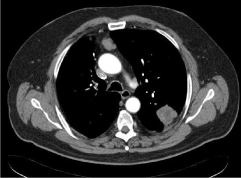
Figure 1. Contrast CT of the chest: left-lung carcinoma and anterior mediastinal thymoma

Figure 2. (A) High-grade squamous cell lung carcinoma showing nests with multilayer arrangement of neoplastic cells without keratinization and comedo-like necrosis in the nest’s centers. (B) Mediastinal type AB thymoma comprising densely packed, elongated, bland-looking epithelial cells forming numerous discrete rosettes (left) and lymphocyte-rich areas (right). Hematoxylin/eosin staining, magnification: x200
The postoperative course was uncomplicated. Based on the lung cancer stage, standard adjuvant chemotherapy was administered (cisplatin 80mg/m2 day 1, vinorelbine 30mg/m2 days 1, 8; every 21 days). Due to the stage of a thymoma and uncertain completeness of its resection adjuvant radiotherapy was administered to the thymus bed with a margin (3D technique; X = 15MeV; fractional dose, 200cGy; total dose, 5600cGy). Systemic treatment was terminated after Cycle 1 following gastrointestinal obstructive symptoms likely related to vinorelbine. After improvement of the patient’s general condition, radiotherapy was conducted as planned. Forty-eight months post-surgery, the patient was in very good general condition, with no clinical or radiological features of tumor progression for either malignancy.
The validity of simultaneous surgical treatment of NSCLC and thymoma has been documented, but published case reports are few and have not involved adjuvant treatment [3].
In this case, following inconclusive bronchoscopy, thoracotomy was selected and enabled adequate surgery (lobectomy with lymphadenectomy). NSCLC diagnosis was confirmed intra-operatively. Based on radiology, thymectomy was selected before the thymic tumor histology had been established. The clinical stages of squamous cell carcinoma and thymoma were post-operatively determined as IIB and Masaoka-Koga III, respectively. According to current TNM staging system for thymic tumors a stage should be assessed as I, however postoperative clinical decisions were based on Masaoka-Koga classification used at the time of primary diagnosis.
Tumor stage is the most important factor determining eligibility for adjuvant therapy for both cancers. In stage II NSCLC, adjuvant cisplatin/vinorelbine significantly increases 5-year overall survival by 11.6% [4]. Systemic adjuvant treatment after radical thymoma resection is not recommended; however, adjuvant radiotherapy is indicated for Masaoka-Koga stage III thymoma and reduces mortality due to thymic tumor recurrence [5-6].
This report documents the management and response of a patient who underwent radical treatment for two concurrent primary thoracic tumors. The low clinical stage of both tumors enabled simultaneous thymectomy and lobectomy with lymphadenectomy. There were no clinically significant perioperative complications. Adjuvant therapy was administered based on guidelines for both tumor types. No clinical or radiological features of relapse were observed after 4 years. We propose this treatment protocol may be safe and effective for other patients with this extremely rare presentation.
- Filosso P, Galassi C, Ruffini E, Margaritora S. Bertolaccini L, et al. (2013) Thymoma and the increases risk of developing extrathymic malignancies: a multicenter study. Eur J Cardiothorac Surg 44: 219-224.
- Kamata T, Yoshida S, Wada H, Fujiwara T, Suzuki H, et al. (2017) Extrathymic malignancies associated with thymoma: a forty-year experience at a single institution. Interact Cardiovasc Thorac Surg 24: 576-581.
- Song X, Shen H, Li J, Wang F (2016) Minimally invasive resection of synchronous triple primary tumors of the esophagus, lung and thymus: A case report. Int J Surg Case Rep 29: 59-62.
- Douillard J, Tribodet H, Auber D, Shepherd FA, Rosell R, et al. (2010) Adjuvant cisplatin and vinorelbine for completely resected non-small cell lung cancer subgroup analysis of the lung adjuvant cisplatin evaluation. J Thorac Oncol 5: 220-228.
- Zhou D, Deng X, Liu Q, Zheng H, Min JX, et al. (2016) The effectiveness of postoperative radiotherapy in patients with completely resected thymoma: a meta-analysis. Ann Thorac Surg 101: 305-310.
- Girard N, Ruffini E, Marx A, Faivre-Finn C, Peters S, et al. (2015) Thymic epithelial tumours: ESMO Clinical Practice Guidelines for diagnosis, treatment and follow-up. Ann Oncol 26 Suppl 5: v40-55.