An 8-year old boy was admitted to our emergency department after falling off his bike. He sustained two frontal skin lacerations and an intraoral wound. 36 hours after the trauma the patient developed seizures and fever, and was readmitted to the emergency unit. The leukocyte count now had increased from 17 to 35/nl and the CRP from 0 to 70 mg/l. All blood and CSF cultures showed heavy growth of Streptococcus pneumoniae. Appropriate antibiotic treatment was started following admission to the paediatric intensive care unit. An initial MR scan did not reveal signs of brain damage but gave rise to the suspicion of a basilar skull fracture through the lamina cribrosa. Soon the clinical signs of brain stem compression developed. A repeat MR scan 7 hours later showed global ischemic brain damage together with compromised brain stem perfusion. An emergency decompression craniectomy of the brainstem and insertion of ventricle drainages was performed. 12 hours later no brain stem reflexes could be elicited and therefore dissociated brain death was diagnosed by repeated clinical examinations.
Here we discuss the dismal course of a non-vaccinated boy subject to overwhelming invasive meningitis by streptococcus pneumoniae secondary to a frontal sinus/basilar skull fracture.
An 8-year old boy was admitted to the emergency department after falling off his mountain bike. His initial Glasgow Coma Score (GCS) was 15/15, there were no focal neurological findings, and he was well orientated. There were two (1 and 2 cm) superficial wounds on his forehead and bruising underneath the right eye and a minor laceration of the lip. The orthopaedic resident on call obtained a medical history that revealed nothing abnormal except from not having received the recommended immunizations. The boy complained of pain around the right eye and vomited once after the initial examination. Routine blood samples were taken, and the planned MRI scan of the brain was cancelled after the family refused the special investigation. The orthopaedic resident then discussed the situation with the paediatrician on-call and it was decided to rather obtain an x-ray of the temporomandibular joints and to refer the patient to maxillofacial surgery for an evaluation of the intraoral wound. No fractures were noted on the plain x-ray of the mandible.
After the maxillofacial surgeon had sutured the facial wounds the patient was admitted to the paediatric ward for ½ hourly neurologic observations. During the night the mother remained with her child. The night staff reported that the child slept well with no complaints of nausea or vomiting. Since admission all nursing observations were normal. The next morning the patient was seen by the senior orthopaedic surgeon. On examination the patient was in a good condition, had passed urine, eaten well and no further medical conditions were reported. A wound check did not show any signs of wound infection or bleeding. Upon discharge the parents were instructed to return immediately or call an emergency service in case of neurologic signs such as nausea, headache, or drowsiness.
At 5 a.m. the next morning the child was admitted via the emergency service in an unresponsive and pyrexial state. According to the mother he had developed a temperature early that morning and started vomiting. He had also complained of neck pain which was interpreted by his mother as positional. The patient then suffered a seizure which the mother decided to treat by a cold-water enema. After initial improvement, the patient became unresponsive and the cramps continued. The emergency service was called.
On initial examination he was found to be unconscious with his upper limbs in internal rotation, his legs were fully extended and spastic. His temperature was 39.8°C. Midazolam was given to stop his seizures. A MRI scan was obtained under sedation which showed no signs of brain injury, but a moderately increased signal around the right bulbus without any sign of optical nerve involvement. A small amount of air was noted in the orbita suggestive of an in-situ fracture of the orbital wall/frontal sinus superiorly or medially (Figure 1). The muscle spasms decreased and he was taken to the paediatric ward for further observation, here he remained unresponsive. Blood results revealed a CRP of 70 mg/L (0 mg/L 36 hours prior), a leukocyte count of 37/nL (16/nL 36 hours prior), and mild hyponatremia (134 mmol/L).
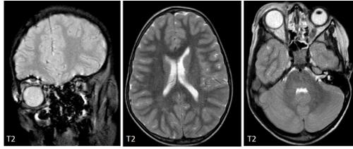
Figure 1. T2 MRI brain and skull performed after re-admission. Odema around the skull base indicates fracture of the lamina cribrosa. The brain is seen without abnormality. No brainstem entrapment is present
After one hour of observations, the mother called for a nurse because suddenly the patient showed agitation, tachycardia (150 bpm), and his oxygen saturation dropped to below 70% on supplementation Oxygen of 4 L/min. The paediatrician on call immediately re-examined the patient, as he found the patient to be apnoeic, he was intubated and ventilated in the paediatric intensive care unit. A loading dose of cefotaxime and tobramycin was administered. The pupils were now unresponsive and of unequal size, so a doppler of the brainstem was performed which showed decreased signaling of the brainstem vascular supply. A repeat MRI of the brain was then taken that now showed hypo-intense white matter changes, missing flow-void of cortical veins with signs of intravascular coagulation, an extremely reduced arterial perfusion, and imminent cerebellar herniation. The imminent herniation was discussed with the neurosurgeon on-call, and after informed consent by the parents, immediate brain stem decompression craniectomy and the insertion of ventricular drainages was performed. CSF analysis demonstrated overwhelming infection not limited to the meninges (leukocytes 13.011/µL, protein >6.000 mg/L, glucose <0.11 mmol/L). The next morning dissociated brain death was diagnosed by lack of brain stem reflexes and electrophysiologic examination, and after a repeated clinical investigation intensive care treatment was withdrawn.
Cultures of blood and of CSF showed rapid growth of Streptococcus pneumoniae. Finally, the serotype 34 was identified which does not form part of any vaccination program given to children.
The dismal course of the invasive pneumococcal infection presented here mainly raises three questions:
- Is the infection related to the head injury the child had suffered almost 60 hours before death;
With the frontal sinuses being patent in our 8 year old patient the demonstration of air within the orbita by MRI provides a rationale for the most likely route of bacterial transmission via a CSF leakage through the lamina cribrosa (Figure 2). Invasive pneumococcal infection is a known complication of basilar skull fractures. In a French national survey assessing children older than 5 years with pneumococcal meningitis any kind of cerebrospinal fluid leakage (primary, traumatic, unknown reasons) caused bacterial transmission in 58% (n=63) of all study participants [1].
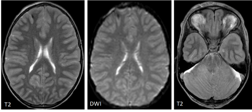
Figure 2. Follow-up MRI taken 6 hours after re-admission. T2 weighted images show diffuse swelling of the white matter. Diffusion weighted images show global diffusion changes in keeping with severe brain damage. The ventricular system is severely obstructed and there is brain stem entrapment
- Was appropriate treatment performed;
Whether antibiotic prophylaxis should be administered in every case remains debatable. There is little evidence to support routine administration in basilar skull fractures as recently reviewed by a Cochrane analysis, however, most of the included trials are more than 20 years old and present data from children and adults of the pre-vaccination era [2]. Early discharge is often advocated when paediatric patients with undisplaced base of skull fracture do not present with neurologic symptoms. An American study exclusively assessing frontal sinus fractures described serious infections in 14/242 (5.8%) while the overall prevalence of CSF leak was 36.1% and 5.4% for more than 7 days, respectively [3]. All patients with serious infections had both involvement of the posterior table of the frontal sinus and nasal outflow tract injury, and perioperative antibiotics beyond 48 hours were not associated with a decreased risk for infection.
As pneumococcal meningitis is known to progress rapidly, administration of cefotaxime 200 mg/kg and tobramycin 5 mg/kg was immediately started upon re-admission. Pneumococcal resistance against cefotaxime is rare in Germany (<1% of meningitis isolates) [4]. Stabilization of organ failure was carried out by internal standards with early intubation, ventilation, sedation and haemodynamic monitoring. Despite appropriate antibiotic treatment approximately 10% of children will die from overwhelming infection [1].
- How may the state of being non-vaccinated be interpreted in our patient with regard to disease progress?
Pneumococci may cause otitis media, pneumonia, meningitis, sepsis and haematogenic osteomyelitis for which reasons a pneumococcal conjugate vaccine (PCV7) was introduced in 2006 to the German guidelines for vaccination, nowadays being expanded to PCV13 covering the serotypes 1, 3, 4, 5, 6A, 6B, 7F, 9V, 14, 18C, 19A, 19F and 23F. Conjugate vaccines (containing serotype-specific polysaccharides conjugated to carrier proteins) induce antibodies that are specific for each serotype (with little cross-reactivity) and that opsonize bacteria followed by phagocytosis [5]. The widespread use of PCV7, however, led to an increase in disease caused by non-vaccine serotypes through a phenomenon called “serotype replacement”. By the introduction of PCV13 the incidence of pneumococcal meningitis could be reduced by 44% as shown in a French cohort comparing the years 2009 and 2014 [6]. In contrast, the immunizing effect of PPSV23 (a polysaccharide vaccine without conjugation to carrier proteins) is T-cell independent, does not induce memory and is therefore not recommended for regular use in infants, but as add-on prophylaxis in immunocompromised conditions or chronic diseases such as congenital heart disease or diabetes.
Neither PCV13 nor PPSV vaccines include the serotype 34 isolated in our patient, suggesting a niche replacement by this less dominant strain. A Portuguese study demonstrated mucosal colonization of the more invasive PCV13 serotypes in 6.4% of the vaccinated group, and in 38.5% of the non-vaccinated group increasing the risk of colonization by the factor 6 in the non-vaccinated group [7]. Overall mucosal colonization with pneumococcus species did not differ between the groups with 66.5% in the vaccinated group and 60.1% in the non-vaccinated group. The advance of non-vaccine serotypes as well as of non-pneumococcal mucosal colonizators such as H. influenza, S. aureus, and M. catarrhalis was also confirmed in a Dutch study enrolling infants and toddlers between 2005 (pre PCV7) and 2013 [8]. Taken together, the state of being non-vaccinated of our patient carries an increased risk for suffering a major invasive infection by e.g. serotype 34, however, in addition disease progress might have been accelerated.
Rapid progression of pneumococcal meningitis/encephalitis to death typically occurs within <72 hours of disease [9]. Risk factors for mortality are respiratory failure, septic shock, multiple organ failure and low CSF leukocyte count (<200/µL), hyponatremia, seizures and focal neurological signs [10,11]. While our patient experienced several of the risk factors mentioned above, his CSF status did not present signs of immunologic hypo-reactivity but of complete failure/destruction of the meningeal functions suggesting meningitis and encephalitis.
Diffusion-weighted imaging (DWI), fluid attenuated inversion recovery (FLAIR), and contrast-enhanced T1-weighted sequences are considered superior in detecting and monitoring complications such as infarction, vasculitis, cerebritis, and intraventricular or subdural empyema. In a cohort of adult patients with proven meningitis by CSF changes, only 82% showed intracranial MRI abnormalities such as intraventricular/sulcal diffusion restriction, and hypointense white matter changes were only detected in 26% [12]. Other meningitis-associated complications such as dilatation of the veins as demonstrated in our patient are rare events [13]. Detection of meningitis in the first MRI investigation was probably missed because the FLAIR sequence was not contrast enhanced (CE). CE-FLAIR was shown to be superior to CE-T1W sequences to detect meningeal enhancement in a cohort of 60 paediatric and adult patients with proven meningitis by CSF [14].
Invasive pneumococcal infection is a potentially life-threatening disease because of its rapid progression to systemic infection involving meningoencephalitis. Basilar skull fractures and frontal sinus fractures are typical traumatic lesions causing CSF leak and bacterial transmission. Despite appropriate antibiotic treatment approximately 10% of paediatric patients demise because of the rapid spread of the infection. This case demonstrates a tragic clinical course of an infection that may have been prevented or attenuated by complying with the German guidelines for vaccination even though the causative serotype 34 must be considered as serotype replacement affecting both, vaccinated and non-vaccinated children. The rapid progression of the meningitis/encephalitis was only detected by clinical signs as MRI failed to show infection-specific findings, probably also due to not considering CE-FLAIR. A case like this should raise concern towards a growing number of opponents of vaccinations as some of the most severe infections can be prevented.
- Henaff F, Levy C, Cohen R, Picard C, Varon E, et al. (2017) Risk factors in children older than 5 years with pneumococcal meningitis: Data from a national network. Pediatr Infect Dis J 36: 457-461. [Crossref]
- Ratilal BO, Costa J, Pappamikail L, Sampaio C (2015) Antibiotic prophylaxis for preventing meningitis in patients with basilar skull fracture. Cochrane Database Syst Rev 28: CD004884. [Crossref]
- Bellamy JL, Molendijk J, Reddy SK, Flores JM, Mundinger GS, et al. (2013) Severe infectious complications following frontal sinus fracture: The impact of operative delay and perioperative antibiotic use. Plast Reconstr Surg 132: 154-162. [Crossref]
- Imohl M, Reinert RR, van der Linden M (2015) Antibiotic susceptibility rates of invasive pneumococci before and after introduction of pneumococcal conjugate vaccination in Germany. Int J Med Microbiol 305: 776-783. [Crossref]
- Miyaji EN, Sarno Oliveira ML, Carvalho E, Ho PL (2013) Serotype-independent pneumococcal vaccines. Cell Mol Life Sci 70: 3303-3326. [Crossref]
- Cohen R, Varon E, Bechet S, Bonacorsi S, Levy C (2016) Comparative impact of pneumococcal conjugate vaccines on pneumococcal meningitis according to underlying conditions. Vaccine 34: 4850-4856. [Crossref]
- Valente C, Hinds J, Gould KA, Pinto FR, de Lencastre H, et al. (2016) Impact of the 13-valent pneumococcal conjugate vaccine on Streptococcus pneumoniae multiple serotype carriage. Vaccine 34: 4072-4078. [Crossref]
- Bosch AATM, van Houten MA, Bruin JP, Wijmenga-Monsuur AJ, Trrzcinski K, et al. (2016) Nasopharyngeal carriage of Streptococcus pneumoniae and other bacteria in the 7th year after implementation of the pneumococcal conjugate vaccine in the Netherlands. Vaccine 34: 531-539. [Crossref]
- Hsiao HJ, Wu CT, Huang JL, Chiu CH, Huang YC, et al. (2015) Clinical features and outcome of invasive pneumococcal disease in a pediatric intensive care unit. BMC Pediatrics 15: 85-90.
- Tsai MH, Chen SH, Hsu CY, Yan DC, Yen MH, et al. (2008) Pneumococcal meningitis in Taiwanese children: Emphasis on clinical outcomes and prognostic factors. J Trop Pediatr 54: 390-394. [Crossref]
- Chao YN, Chiu NC, Huang FY (2008) Clinical features and prognostic factors in childhood pneumococcal meningitis. J Microbiol Immunol Infect 41: 48-53. [Crossref]
- Lummel N, Koch M, Klein M, Pfister HW, Bruckmann H, et al. (2016) Spectrum and prevalence of pathological intracranial magnetic resonance imaging findings in acute bacterial meningitis. Clin Neuroradiol 26: 159-167. [Crossref]
- Mukherjee D, Saha A (2017) Cerebral vasculitis in a case of meningitis. Iran J Child Neurol 11: 81-84. [Crossref]
- Azad R, Tayal M, Azad S, Sharma G, Srivastava RK (2017) Qualitative and quantitative comparison of contrast-enhanced fluid-attenuated inversion recovery, magnetization transfer spin echo, and fat-saturation T1-weighted sequences in infectious meningitis. Korean J Radiol 18: 973-982. [Crossref]