The orthodontic treatment of impacted maxillary canine remains a challenge to today's clinicians. The treatment of such clinical cases usually involves surgical exposure of the impacted tooth, followed by orthodontic traction to guide and align it into the dental arch. Bone loss, root resorption, and gingival recession around the treated teeth are some of the most common complications.
The main concern of the orthodontist during designing the treatment plan is the anchorage. The raising of the (TAD) will help a lot in canine traction in the proper direction without any impairment on the adjacent tissues.
In this article, a case report of maxillary labially impacted canine which had been guided to its position via two surgical approaches with the assist of micro implant.
impacted, canine, temporary anchorage device (TAD)
Impacted tooth is one that fails to erupt and will not attain its anatomical position beyond the chronological eruption date even after its root completion. The impacted maxillary canine is a frequently encountered clinical problem. Maxillary canine is the most commonly impacted teeth, second only to third molars [1]. The permanent maxillary canine is frequently misplaced teeth in relation to other teeth in the maxilla. The canine is the most frequently found palatal to the lateral incisor. Impactions are twice as common in females. 8% have bilateral impactions [2]. Approximately, one-third of impacted maxillary canine is located labially, and two-thirds are located palatally [3].
Labially impacted canine is mostly due to tooth size-arch length deficiency. Study shows only 17% of labial impacted canine shows insufficient space, whereas palatally impacted canine has sufficient space in 85% cases for eruption [4].
Upper right labially impacted canine impaired the patient smile who came to the private clinic seeking for a solution. The humble request of the patient not to extract any of the permanent teeth in order to solve her problem. On examination found that #13 is overlapping the apical 1/3 of the lateral incisor. The treatment plan designed to disimpact and traction of the canine using a microimplant through two surgical stages. This article illustrates the step-by-step of orthodontic and surgical procedures in efficiently recovering labially impacted maxillary canine.
A 12-year-old female came to the dental clinic in the private sector with a chief complaint of unpleasant smile as the upper lateral incisor is flared and missed upper right canine. During our discussion with the patient, her humble request was not to lose any of the permanent teeth. On clinical examination, it’s found that #12 is labially proclined, retained upper right primary canine with slight soft tissue elevation in the labial sulcus on palpation. Intraoral photographs showed misalignment of teeth with shifted upper midline to the right side (Figure 1). A poor oral hygiene had been noticed with a generalized mild upper and lower gingivitis. Panoramic radiograph showed impacted #13 which is overlapping the apical 1/3 of the lateral incisor with retained primary canine (Figure 2). The mesial surface of the canine is passing the root of the lateral and almost flushing with distal surface of the apical third of the central incisor. Periapical radiograph in different horizontal directions (SLOB) techniques along with the palpation and the labial proclination of the lateral incisor confirmed that the impacted canine is labially located. Lateral cephalogram revealed that the incisal edge of the canine is labial to the roots of the incisors. Cephalography analysis documented that patient has skeletal class I malocclusion with decreased anterior facial height and relatively proclined upper and lower incisors. The soft tissue analysis showed that upper and lower lips are relatively behind the Esthetic line (E-line) by 4mm for the upper and 3.6mm for the lower (Table 1).
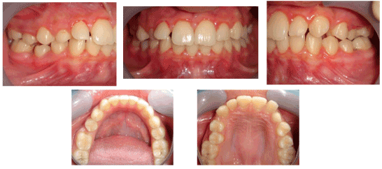
Figure 1. Pre-orthodontic intra oral digital photos
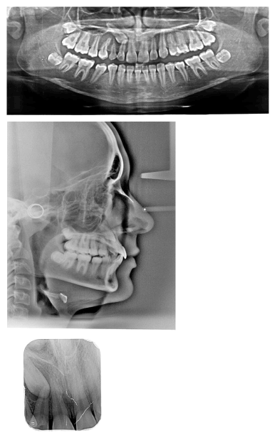
Figure 2. Pre-orthodontic intra oral digital photos
Table 1. Cephalometric analysis
|
Parameter |
Normal Value |
Pre-treatment |
Post treatment |
Sagittal relations |
|
|
|
SNA |
82°±2 |
78° |
78.4° |
SNB |
80°±2 |
77.3° |
78° |
ANB |
0-4° |
+0.7° |
+0.4° |
Wits’ |
-1mm/ 0 mm |
|
|
SN-Ba |
130° |
140.5° |
142.1° |
SN- Pog |
81° |
78.9° |
78.2° |
Vertical relations |
|
|
|
SN/FH |
5-7° |
10.1° |
10.7° |
SN-Max pl |
9° |
7.6° |
8.3° |
SN- Mand pl |
32° |
25.8° |
25.4° |
Mp- Max. interbasal ang. |
24° |
16.9° |
16.4° |
PFH/AFH (Jarabak ratio) |
59-62% |
69.2% |
65.7% |
Gonial angle |
120° |
108.7° |
112.1° |
Y axis angle FH/S-Gn |
59.4° |
|
|
Dental Analysis |
|
|
|
U1/ SN |
102° |
107.6°
|
111.1° |
U1/ Max pl |
109° |
115.5° |
120.2° |
U1/ NA |
22° |
30.9° |
33.8° |
U1/NA |
+4 mm
|
+11.2mm
|
+6.2mm
|
L1/Mp
|
90°
|
106.3°
|
100.9°
|
L1/NB |
25° |
28.6° |
24.2° |
L1/NB |
+4 mm
|
+7.4mm
|
+3.3mm
|
L1/A-Pog |
22° |
31.9° |
27.4° |
L1/A-Pg |
1 mm |
+4mm |
+3mm |
Interinc.angle |
135° |
119.8° |
122.4° |
Soft tissue analysis |
|
|
|
Up Lip/E line
|
-4 mm
|
-7mm
|
-2.6mm
|
Lo Lip/ E line
|
-2 mm
|
-5.6mm
|
-2.2mm
|
Nasolabial angle |
102°±4
|
100° |
103.9° |
Labiomental angle |
120° |
120° |
117.3° |
Z angle |
80°±9 |
80.2° |
80.2° |
Angle of facial convexity |
12°±4 |
8.8° |
7° |
Maxilla (all three planes):
- A - P: Maintain
- Vertical: Allow for normal expression of growth
- Transverse: Maintain
Mandible (all three planes):
- A - P: Allow for normal expression of growth
- Vertical: Allow for normal expression of growth
- Transverse: Maintain
Maxillary Dentition
- A - P: retroclination
- Vertical: Maintain
- Inter-molar Width: Maintain
Mandibular Dentition
- A - P: retroclination
- Vertical: Maintain
- Inter-molar / Inter-canine Width: Maintain
Facial Esthetics:
- Upper/lower lips should be in more forward position in relation with E line.
Non-extraction treatment with a full fixed orthodontic appliance was indicated to align and level the dentition. In the initial stage of treatment, space was opened for the impacted canine and the patient was referred for the first stage of surgical exposure. The vertical incision subperiosteal tunnel access (VISTA) technique was selected due to the relatively high, horizontal position of the impacted tooth (above MGJ). After crown exposure, a button had been placed and the horizontal traction anchorage started directly on extra- alveolar microimplant which inserted in the infrazygomatic crest parallel to the upper right first molar. The traction activated by subsequent adjustment of the power chain (PC) in order to move the tooth under its socket. The patient referred for the 2nd stage of surgery in order to begin with the vertical traction where the PC directly connected to the main arch wire. Fixed appliances were removed, and the corrected dentition was retained with clear overlay on both arches.
0.022 slot Roth brackets had been bonded on all upper and lower teeth except #12 which acted as a free body initially till the canine had been moved away from its root (Figure 3). After 6 months of alignment, space started to be created for the canine using opening coil spring (0.010 x 0.035 inch) (Figure 4). The first stage of surgery was planned to be VISTA technique in order to move the canine horizontally and to situate the canine crown directly under its socket using the microimplant (MI) (Figures 5 and 6). During surgery, all the bone distal to the canine crown till its CEJ which is in the way of its movement had been removed. A microimplant from (Ormco) VectorTas of 2 x 8 mm had been placed in the infrazygomatic crest parallel to the upper right first molar (Figure 7). A lingual button bonded on the labial surface of the canine and connected to the microimplant via a power chain which was passing under the alveolar mucosa over the canine. The horizontal movement of the crown had been activated every month by cutting a hole from the power chain. A panoramic periapical radiograph A-B taken directly after MI placement and 3 months over that to control the movement of the canine (Figures 8 and 9). After 3 months of horizontal movement of the canine, the patient referred again for the 2nd stage of surgery where a full reflected flap performed in order to remove the old power chain and place new one which is directly connected to the main archwire (Figure 10). All the bone above the canine crown till the 2 mm from the alveolar crest had been removed in order to facilitate the tooth movement vertically. The main arch wire which is 0.017x0.025 Stst had been offset in the area between #12, 14 (Figure 11). This offset placed to help for keeping the canine root in the alveolar bone and avoid the labial tipping of the crown. A crimpable attachment with a hook fixed on the wire directly over the canine crown and a power chain connected directly from the lingual button to the hook (Figure 12). The vertical movement of the crown had been activated every month by cutting a hole from the power chain. After the canine came out of the soft tissue a bracket bonded and thin wire placed in its slot with a sequence of 0.12 Niti, 0.14 Niti, 0.16 Niti, 0.16 x 0.22 Niti, 0.16 x 0.22 Stst, 0.17 x 0.25 Niti and 0.17 x 0.25 Stst. Canine root torque had been checked after its reaching to the occlusal plane and found that no need for any adjustment since it is similar with the opposing canine root eminence (Figure 13). After 24 months of active treatment, all appliances were debonded (Figure 14). Orthopantogram, lateral cephalography and periapical radiograph had been taken to record as a baseline for future follow up and assessment. Clear overlays delivered for both arches as retainers with proper instructions (Figure 15).

Figure 3. Intra oral photos of the initial visits of levelling and alignment

Figure 4. Intra oral photos of the stage of creating space for the impacted #13

Figure 5. Photos show the placement of microimpant in the region of #16 and using power chain to move #13 distally in the horizontal plane
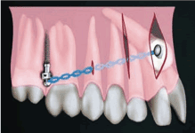
Figure 6. A schematic drawing of the recently developed VISTA technique (Vertical Incision Subperiosteal Tunnel Access), a minimally technique, combined with bone screws which is indicated in labially impacted cuspids
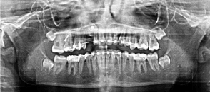
Figure 7. A panoramic radiograph shows the insertion of the micro implant and the button which had been to the labial surface of #13
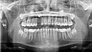
Figure 8. A panoramic radiograph 3 months past traction #13
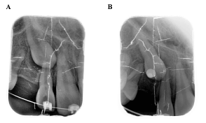
Figure 9. A- Intra oral radiograph immediately before horizontal movement of #13; B-3 months post traction #13
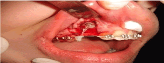
Figure 10. Surgical procedure of bilateral vertical release and vertical movement of #13 via using a power chain

Figure 11. Intra oral photos show the vertical traction #13
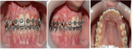
Figure 12. Intra oral photos with a button which attached on the canine to be shown through the soft tissue as #13 started to erupt
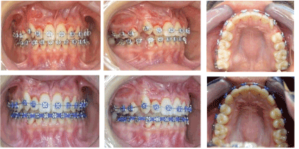
Figure 13. Bracket bonded on #13 after its eruption and starting with its levelling and alignment

Figure 14. Intra oral photos of post orthodontic treatment
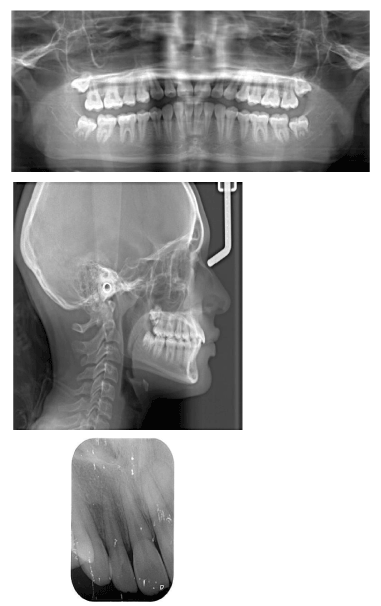
Figure 15. Panoramic, lateral cephalometric and peri apical radiographs after orthodontic treatment finished
The etiology of labially impacted canine is often attributed to abnormal position of tooth buds, associated with an arch length and/or width deficiency. The three methods for diagnosing impacted canines are inspection, palpation, and radiography. Inspection and intraoral palpation of the canine bulge are useful for determining the general location of the impacted canine [5]. However, three-dimensional cone beam computed tomography (CBCT) images are the standard of care for providing the most accurate information about the location of the impaction relative to its adjacent teeth.
The treatment modalities of labially impacted canine starts with non-extraction treatment which is indicated unless there are other complications, such as severe crowding, ankylosis, uncontrolled infection, internal or external root resorption, severe root dilacerations, and/or pathology that may compromise adjacent teeth during or after orthodontic treatment [6].
Non-surgical approach: According to Williams study in 1981, selective removal of deciduous cuspids is a suggested interceptive measure in Class I uncrowded malocclusions [7]. Olive concluded that creating space for the impacted canine with fixed appliances, and waiting for spontaneous eruption, is an effective option. Many impacted canines cannot be treated with nonsurgical methods [8]. If impacted canines do not erupt after a year of treatment, then surgical intervention is indicated. Kokich summarized three techniques for treating labially impacted maxillary canines, including excisional uncovering (E), apical positioned flap (APF), and the closed eruption technique (CE) [9]. Their indications and contraindications are shown in (Table 2).
Table 2. Surgical considerations for labially impacted cuspids
|
E |
APF |
CE |
B-L position (if surrounding bone wrapped the surface of crown) |
x |
x |
✓ |
Crown apical to MGJ |
x |
✓ |
✓ |
The mount of attached gingiva < 2-3mm |
x |
✓ |
x |
M-D position (if the crown’s position overlapped with the root of lateral incisor) |
x |
✓ |
x |
E: excisional uncovering; APF: apically positioned flap; CE: closed eruption technique; MGJ: mucogingical junction
In the present case, upper right impacted canine was tilted mesially and positioned across the middle of the root of the adjacent lateral incisor. For less scar formation, particularly in the esthetic zone, the Vertical Incision Subperiosteal Tunnel Access (VISTA) technique, provides minimally invasive alternative for the surgical treatment of labial impactions [10, 11].
The assist of micoimplant was very successful and helpful to move the impacted canine away from the root of the lateral incisor in order to avoid any root resorption. The treatment plan should be designed before orthodontic treatment starts in order to know which bone should be removed to facilitate the impacted tooth movement in both directions horizontally and vertically.
The proper case evaluation for labially impacted canines using VISTA technique and TADs will treat many of such cases which had been extracted previously and comply the request of most of the patient which is that does not like to have any permanent tooth to be missed.
- Mesotten K, Naert I, van Steenberghe D, Willems G (2005) Bilaterally impacted maxillary canines and multiple missing teeth: A challenging adult case. Orthod Craniofac Res 8: 29-40. [Crossref]
- Bishara SE (1992 ) Impacted maxillary canines: A review. Am J Orthod Dentofacial Orthop 101: 159-171. [Crossref]
- Ericson S, Kurol J (1986) Radiographic assessment of maxillary canine eruption in children with clinical signs of eruption disturbance. Eur J Orthod 8: 133-140. [Crossref]
- Jacoby H (1983) The etiology of maxillary canine impactions. Am J Orthod 84: 125-132. [Crossref]
- Richardson G, Russell KA (2000) A Review of impacted permanent maxillary cuspids-Diagnosis and prevention. J Can Dent Assoc 66: 497-501. [Crossref]
- Tseng SP, Chang CH, Roberts WE (2009) High maxillary canine impaction with mesial and labial displacement-ABO case report. News & Trends in Orthodontics 18: 36-44.
- Williams B (1981) Diagnosis and prevention of maxillary cuspid impaction. Angle Orthod 51: 30-40. [Crossref]
- Leite HR, Oliveira GS, Brito HH (2005) Labially displaced ectopically erupting maxillary permanent canine: interceptive treatment and long-term results. Am J Orthod Dentofacial Orthop 128: 241-251. [Crossref]
- Kokich VG (2004) Surgical and orthodontic management of impacted maxillary canine. Am J Orthod Dentofacial Orthop 126: 278-283.
- Su B, Hsu YL, Chang CH, Roberts WE (2011) Soft Tissue Considerations for the management of Impactions. Int J Orthod Implantol 24: 50-59.
- Hsu YL, Chang CH, Roberts WE (2011) A closed eruption technique modified from vertical incision subperiosteal tunnel access VISTA. Int J Orthod Implantol 24: 60-67.


