A young female patient of 15y.o presented at my hospital by dyspnea on effort and palpitation for one year. Mental deficiency was notified. Physical examination detected a 3/6 continuous murmur at the 2ndRICS. In the past history, PDA had been suspected by her physician, associated with recurrent bronchitis.
Trans-thoracic Echocardiography showed an enlarged LV of 57mm with normal EF of 69%, LCA=5mm, RCA=3.5mm at origin, no suspected sign of PDA was seen. Only a continuous flow was visualized in the PA.
CT-Angiography with IV contrast medium showed the Left Common Carotid Artery rising from the Pulmonary Artery trunk. PDA was not presented.
The Left Common Carotid Artery then was re-implanted into the aortic arch normally with a favourable post-operative progress
Carotid artery, Pulmonary artery, Anomalous origin
Anomalous origin of the left common carotid artery is very rare and has been reported previously. We present an operated case of this topic with clinical finding, cardiac ultrasound and MDCT imaging.
A young female patient of 15y.o presented at my hospital by dyspnea and palpitation when running and fast walking for one year. Mental deficiency was notified, she had some difficulties to learn at school. Physical examination detected a 3/6 continuous murmur at the 2ndRICS. In the past history, PDA has been suspected by her physician, associated with recurrent bronchitis. Her body state was normal with 1m60 of height and 48 kg of weight.
She was evaluated immediately by a chest X ray that showed a right aortic arch (Figure 1).
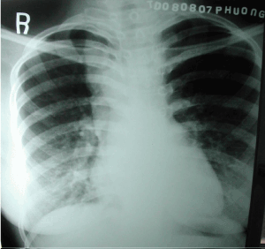
Figure 1. Right aortic Arch.
The trans-thoracic echocardiography that revealed an enlarged LV of 57mm with normal EF of 69% (Figure 2), LCA=5mm, RCA=3.5mm at origin (Figure 3).
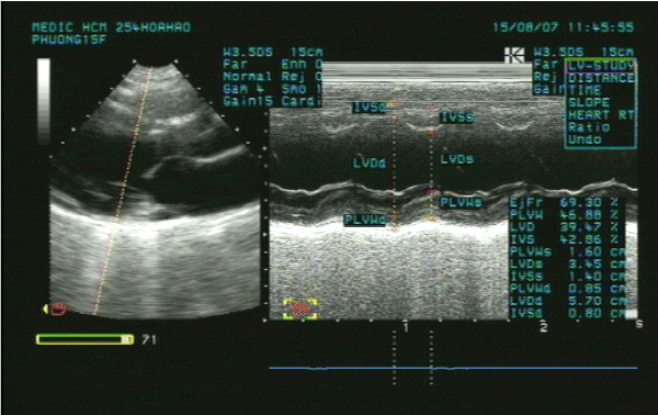
Figure 2. Enlarged LV& normal systolic function.
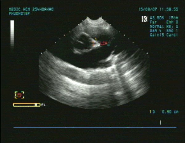
Figure 3. Normal LCA at origin.
No suspected sign of PDA was detected except a continuous flow presented in the Pulmonary Artery (Figure 4).
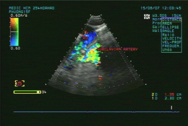
Figure 4. Continuous flow in the PA.
CT-Angiography (MDCT 64) with IV contrast medium Ultravist, slice thickness=1mm visualized a right aortic arch, aberrant origin of the left subclavian artery, dilatation of the branches rising from aortic arch with increased collateral vessels (Figure 5).
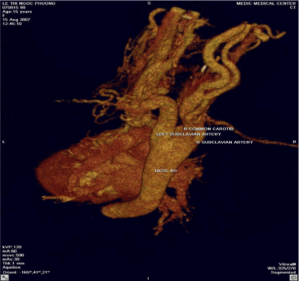
Figure 5. Absence of aortic origin of the LCCA.
Especially, MDCT 64 showed the Left Common Carotid Artery ( LCCA ) rose from the PA trunk (Figure 6). PDA was not detected.
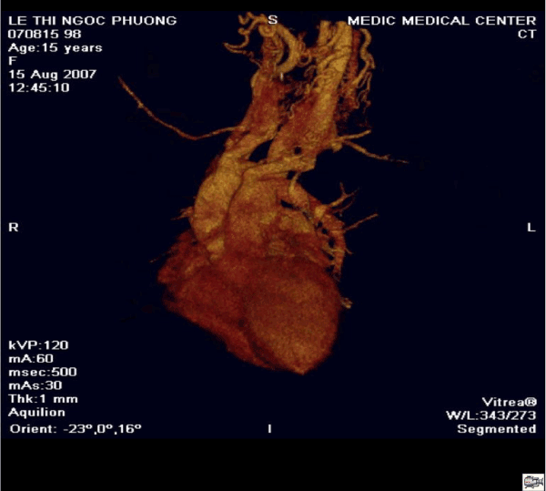
Figure 6. The LCCA rising from the Main PA roof.
Patient underwent uncomplicated surgical repair: the Left Common Carotid Artery was re-implanted into the aortic arch normally with a favorable post-operative progress [1] (Figure 7).
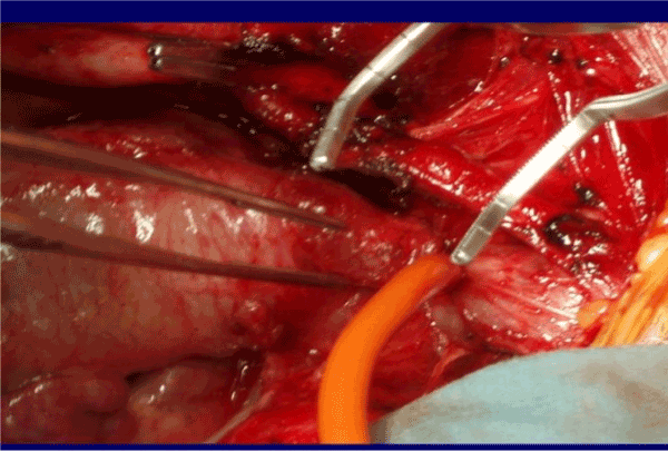
Figure 7. Re-implantation of the LCCA.
Anomalous origin of the Left Common Carotid Artery from the Pulmonary Artery Trunk has been previously reported as rare cases.
Kagami Mijaji has reported a case of anomalous origin of the Innominate Artery from the Right Pulmonary Artery [2].
Onyekachukwu et al. has described a case of anomalous origin of the Left Common Carotid Artery from the Main Pulmonary Artery [3].
Other reports related to the anomalous origin of the Carotid Artery or the Vertebral Artery from a vessel in the systemic circulation like the brachiocephalic trunk.
In this article, my patient was not infant with CHARGES syndrome that includes multiple congenital anomalies like the patients in their reports. She was a teenage patient without other congenital disease.
The role of ultrasound is orienting for the indication of Computed Tomography or DSA. In case of present turbulent flow in the PA, Coronary Fistula and other shunts from the head and neck vessels should be considered.
Anomalous origin of the Left Common Carotid Artery is very rare congenital defect that maybe isolated or associated with some syndromes.
Non-invasive diagnostic methods as Ultrasound and CTA may confirm the diagnosis and inform the anatomical relation of the anomalous vessels prior to operate.
- Hoffmann MHK (2005) Coronary Angiography With Multislice Computed Tomography. JAMA 293: 2371- 2478.
- Miyaji K (2001) Anomalous origin of innominate artery from right pulmonary artery in DiGeorge syndrome. Ann Thorac Surg 71: 2043-2044.
- Osakwe O, Jones B, Hirsch R (2016) Anomalous Origin of the Left Common Carotid Artery from the Main Pulmonary Artery: A Rare Association in an Infant with CHARGE Syndrome. Case Reports in Pediatrics.