Introduction: Determine prevalence of nuchal cords during nuchal translucency screens (NTS), and whether they are associated with adverse perinatal outcomes.
Study design: Retrospective cohort. Study period was 2011 through 2012. NTS images were reviewed by two authors, blinded to pregnancy outcomes. Patients were classified as having nuchal cord present if the cord was documented in grey scale and color doppler. Intrapartum fetal heart rate tracings were classified by NICHD guidelines. Maternal and newborn data were abstracted.
Results: 519 patients had NTS results and obstetrical data available. The prevalence of a nuchal cord during the NTS was 5.7%. There were no statistically significant differences in birth weight, placental insufficiency, and gestational age or mode of delivery, between the two groups. Nuchal cord cases had greater absolute NT measurement (1.7mm vs. 1.59mm; p < 0.001) and abruption diagnosed at delivery (7.4% vs. 0.8%; P=0.035). Nuchal cords at NTS were more likely to be present at birth (40.7% vs. 28.8%; p=0.197).
Discussion: Prevalence of nuchal cord during NTS is 5.7%, with no significant differences in perinatal outcomes, supporting current non-reporting of nuchal cords noted during NTS.
Nuchal cords, nuchal translucency screen, adverse perinatal outcomes, ultrasound, color Doppler
A nuchal cord is defined when the umbilical cord encircles the fetal neck. The finding of nuchal cords at delivery are a rather common phenomenon reported at 16 – 30% [1]. There remains the question of clinical relevance of nuchal cords noted by sonography in the second or third trimesters and if there are any significant, subsequent perinatal adverse outcomes. Several studies have reported on the presence of nuchal cords at delivery; however, there is disagreement in the literature whether there is any impact on birth weight, abnormal intrapartum fetal heart rate tracings, cord gases, Apgar scores and mode of delivery [2-4]. Whether to search for their presence, and then to report their existence on the sonogram report, is still debatable and is imaging-center dependent [5,6]. However, during the nuchal translucency screen (NTS) nuchal cords are searched for, as this alters the nuchal translucency (NT) measurement. There have been two studies [7,8] describing the prevalence of nuchal cords found during the first trimester NTS and the impact the presence of a nuchal cord has on the NT measurement. However, neither study followed their cohort through delivery and perinatal outcomes were not reported, which is the primary focus and aim of our study.
The fetal nuchal translucency thickness is measured between 11 2/7 and 13 6/7 weeks, and it is used in combination with maternal age, maternal serum-free, beta-human chorionic gonadotropin and pregnancy-associated plasma protein-A for aneuploidy screening. The detection rate of chromosomal defects is quoted to be 85-90% [9]. When a nuchal cord is detected in 2D and color Doppler, the NT measurement is made superior and inferior to the nuchal cord, and the two NT measurements are averaged [10].
The primary objective of this study is to report the prevalence of nuchal cords at the time of NTS. Our aim is to describe if there are any associated adverse perinatal outcomes when a nuchal cord is detected at the time of NTS. Our hypothesis is that there is an increase in adverse perinatal outcomes with the presence of a nuchal cord detected on first trimester ultrasounds during the nuchal translucency screen.
This retrospective cohort study was approved for a waiver of consent by the Institutional Review Board at Trihealth, which includes both Good Samaritan Hospital and Bethesda North Hospital in Cincinnati, OH, USA. The list of patient names with singleton pregnancies that were scheduled for nuchal translucency screen from January 2011 to December 2012 was obtained from our genetic counselors. All patients have genetic counseling prior to having the nuchal translucency performed at 11 2/7 to 13 6/7 weeks. All sonographers are RDMS and credentialed in NTS through either the Fetal Medicine Foundation or Nuchal Translucency Quality Review with ongoing monitoring. All the nuchal translucency measurements were interpreted by maternal-fetal medicine specialists. Only the exams that were submitted with a NT measurement for NTS were eligible.
The patient’s demographic, obstetrical, delivery, fetal, and newborn data were obtained from the medical records. Past obstetric history that can impact subsequent perinatal outcomes was reviewed. The data collection sheets were completed with pregnancy and delivery data through the electronic medical record (EMR) systems OBTV (Phillips, Netherlands) in 2011 and then from EPIC (Madison, Wisconsin) from June 2012.
This study was a retrospective review of first trimester ultrasound images in R4 (Hyland Software Inc. West Lake OH), TriHealth sonogram reporting system. To determine if the patient had a nuchal cord at the time of her NTS screen, two of the authors (DL and AK) reviewed all of the sonographic images independently, and then simultaneously, to obtain agreement for each NTS study to determine if a nuchal cord was present or absent. All of the sonographic images for the patients were reviewed with approximately 20 images reviewed per patient study. Both transverse and sagittal images of the fetal neck had been acquired by the sonographers and the umbilical cord position was confirmed by color Doppler. When a nuchal cord was detected in 2D and color Doppler, the NT measurement is made superior and inferior to the nuchal cord and then averaged A patient was classified as a nuchal cord present if the cord was documented in color Doppler and the image had two NT measurements that were averaged for the nuchal translucency measurement. Patients were all de-identified on the data collections sheets. The obstetrical and delivery data were not available to the authors (DL and AK) when reviewing the sonogram images for the determination of nuchal cord presence. All the fetal heart tracings intrapartum were reviewed by AK, without knowledge of nuchal cord presence, and defined by the NICHD fetal heart monitoring guidelines [11].
Since placental insufficiency is not well coded in the EMR, we defined placental insufficiency if any of the following were present: Intrauterine growth restriction (IUGR), low birth weight (LBW), oligohydramnios, or abnormal umbilical artery Doppler (elevated S/D, absent-end diastolic flow or reverse flow).
The investigators suspected the prevalence of nuchal cords at the NTS would be 5% based on prior studies from Schaefer et al. [7] and Scheier et al. [8] A sample size of 456 would allow the study to detect the prevalence with 2% precision and 95% confidence. The two-year study time period included a sufficient number of eligible patients to reach this level of precision. Differences in demographic and clinical characteristics and outcomes between those with a nuchal cord, and those without a nuchal cord, were assessed. Normal distributions of continuous variables were tested; nonparametric statistics were applied where appropriate. Categorical variables were compared using Person’s chi-squared tests, or Fisher’s exact tests, as appropriate. Continuous variables were compared using Student’s t-tests or Mann-Whitney U tests.
Study data were managed using REDCap electronic data capture tools hosted at TriHealth [12]. The statistical analysis was done using IBM SPSS Statistics, Version 21 (SPSS Inc, Chicago, IL).
Of the 658 women who had NTS completed, a total of 519 patients were included in this study. One hundred thirty-nine patients either had no maternal, pregnancy or delivery data or had no nuchal translucency ultrasound available. The patient flowsheet from eligibility, screening to analysis is presented in Figure 1. A total of 35 nuchal cords were found out of 612 patients giving a prevalence of 5.7% of NC found on NTS. Maternal demographic data is shown in Table 1. The groups were well matched without any baseline characteristics differences. While the gestational age at NTS measurement was not different between nuchal cords 12.8 ± 0.67 and no nuchal cords 12.62 ± 0.65 weeks, a significant finding was noted in that in those pregnancies with nuchal cord, the NT measurement was larger at 1.7 mm vs. 1.59 mm (p<0.001).
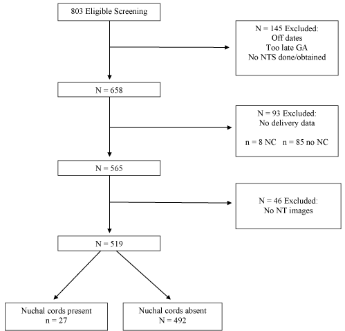
Figure 1. Patient flowsheet from screening to analysis.
Table 1. Demographics, medical and pregnancy history.
| |
|
Nuchal Cord |
No Nuchal Cord |
|
| |
|
mean (SD) |
P* |
Maternal age |
|
32.42 (6.37) |
31.14 (5.89) |
0.302 |
|
|
Median (IQR) |
P† |
Gravidity |
|
2.00 (2.00) |
2.00 (2.00) |
0.925 |
Parity |
|
1.00 (2.00) |
1.00 (1.00) |
0.932 |
Maternal BMI at first visit |
|
25.50 (4.60) |
25.60 (5.70) |
0.483 |
| |
|
N (%) |
P‡ |
Race |
White |
19 (70.4) |
326 (66.3) |
0.525 |
| |
African American |
7 (25.9) |
102 (20.7) |
|
| |
Hispanic |
0 |
12 (2.4) |
|
| |
Other |
1 (3.7) |
52 (10.6) |
|
Chronic hypertension |
|
1 (3.7) |
33 (6.8) |
>0.999 |
Type I or II diabetes |
|
0 |
13 (2.7) |
>0.999 |
Gestational diabetes |
|
2 (7.7) |
55 (11.2) |
0.756 |
Tobacco use |
|
1 (4.0) |
18 (3.8) |
>0.999 |
Previous IUGR |
|
0 |
8 (1.7) |
>0.999 |
APA syndrome/History of thrombosis |
1 (3.7) |
11 (2.2) |
0.479 |
*t-test
†Mann-Whitney U test
‡Chi-squared test
The current pregnancy complications in each group are listed in Table 2. Out of the 27 nuchal cord patients, 2 patients had abruptions, while only 4 of the controls had abruptions (7.4% vs. 0.8%, respectively; p=0.035). There were no differences in preeclampsia, intrauterine growth restriction, decreased fetal movements, polyhydramnios, or oligohydramnios. Given the small number of cases found, most of these complications were not present in the NC patients. Intrapartum fetal heart rate tracing is demonstrated in Table 3. The variable decelerations were present in a higher percentage of the NC patients (51.9% vs. 37.7%; p=0.157). There were no differences in late decelerations, variability of tracing, or prolonged decelerations.
Table 2. Current pregnancy complications.
| |
|
Nuchal Cord |
No Nuchal Cord |
|
| |
|
N (%) |
P* |
Preeclampsia |
|
2 (7.4) |
29 (5.9) |
0.672 |
IUGR |
|
1 (3.7) |
7 (1.4) |
0.353 |
Decreased fetal movements |
0 |
17 (3.5) |
>0.999 |
Any fetal anomalies detected during pregnancy |
0 |
14 (2.9) |
>0.999 |
Polyhydramnios |
|
0 |
13 (2.7) |
>0.999 |
Oligohydramnios |
|
0 |
15 (3.1) |
>0.999 |
Abnormal Dopplers |
|
0 |
5 (1.0) |
>0.999 |
| |
Elevated S/D |
0 |
1 (20.0) |
|
| |
AEDF |
0 |
2 (40.0) |
|
| |
REDF |
0 |
2 (40.0) |
|
Abruption |
|
2 (7.4) |
4 (0.8) |
0.035 |
Placental insufficiency |
|
3 (11.1) |
43 (9.0) |
0.727 |
*Chi-squared test
Table 3. Fetal heart tracing data.
Table 3. Fetal Heart Tracing Data |
| |
|
Nuchal Cord |
No Nuchal Cord |
|
| |
|
N (%) |
P* |
Variable decelerations noted |
14 (51.9) |
183 (37.7) |
0.157 |
Category 3 tracing |
|
0 |
4 (0.8) |
>0.999 |
Late decelerations noted |
|
4 (15.4) |
60 (12.4) |
0.554 |
Variability of tracing |
|
|
|
0.757 |
| |
Minimal |
0 |
5 (1.0) |
|
| |
Moderate |
26 (100) |
467 (97.9) |
|
| |
Marked |
0 |
5 (1.0) |
|
Marked |
|
2 (7.4) |
26 (5.4) |
0.656 |
*Chi-squared test
Delivery data is seen in Table 4. There were no significant differences in average gestational age at delivery, birth weight, Apgar scores, type of delivery, low birth weight, number of vessels, or fetal presentation. Although not significant, a higher percentage of nuchal cords at NTS had nuchal cords present at delivery, as compared with absent nuchal cords at NTS (40.7% vs. 28.8%; p=0.197).
Table 4. Delivery data.
| |
|
Nuchal Cord |
No Nuchal Cord |
|
| |
|
median (IQR) |
P* |
GA at birth |
|
39.14 (2.14) |
39.14 (1.64) |
0.855 |
Birth weight (g) |
|
3360 (400) |
3305 (705) |
0.778 |
Apgar Score – 1 minute |
|
9 (1) |
9 (1) |
0.769 |
Apgar Score – 5 minute |
|
9 (0) |
9 (0) |
0.687 |
| |
|
N (%) |
P† |
Type of delivery |
|
|
|
0.334 |
| |
Spontaneous vaginal |
17 (63.0) |
274 (55.9) |
|
| |
Forceps/vacuum extraction |
3 (11.1) |
24 (4.9) |
|
| |
Cesarean |
7 (25.9) |
192 (39.2) |
|
| |
Reason for cesarean
(multiple responses allowed) |
|
|
|
| |
Nonreassuring FHR |
3 (42.9) |
31 (16.1) |
0.098 |
| |
CPD |
0 |
13 (6.8) |
>0.999 |
| |
Malpresentation |
1 (14.3) |
16 (8.3) |
0.47 |
| |
Failed induction |
2 (28.6) |
43 (22.4) |
0.657 |
| |
Prior C/S |
1 (14.3) |
69 (35.9) |
0.425 |
Malpresentation |
|
1 (3.8) |
22 (4.5) |
0.967 |
Low birth weight |
|
3 (11.1) |
43 (8.8) |
0.724 |
Stillbirth |
|
0 |
9 (1.8) |
>0.999 |
Nuchal cord at birth |
|
11 (40.7) |
141 (28.8) |
0.197 |
| |
Number of loops |
|
|
0.364 |
| |
1 |
11 (100.00) |
118 (84.3) |
|
| |
2 |
0 |
19 (13.6) |
|
| |
3 |
0 |
3 (2.1) |
|
Body cord |
|
1 (4.3) |
15 (3.1) |
0.57 |
Vessels in cord |
2 |
1 (3.7) |
3 (0.6) |
|
| |
3 |
26 (96.3) |
472 (99.4) |
|
Fetal anomaly |
|
0 |
6 (1.2) |
>0.999 |
†Mann-Whitney U test
*Chi-squared test
There were no differences noted in the newborn data, including intubation after delivery, meconium stained fluid, and admissions to the NICU (data not shown).
The primary aim of this study was to establish the prevalence of nuchal cords that are seen on nuchal translucency screen. This was noted to be 5.7%. The secondary aim of this study was to identify any adverse perinatal outcomes that were associated with the finding of NC on the NTS. Similar to published reports [2-4] regarding nuchal cords detected in the second and third trimester, we found no significant differences in the two groups with respect to birth weight, mode of delivery, placental insufficiency or abnormal umbilical artery Doppler. However, the prevalence of these conditions was lower than expected, and the study was underpowered to detect the actual level of difference seen in these results (15% power to detect difference in rate of IUGR; 7% power to detect difference in rate of low birth weight and placental insufficiency).
Placental abruption occurred more frequently in the cases compared to controls (7.4% vs. 0.8%; p =0.035). However, this study was underpowered for this parameter. Several epidemiologic studies have noted that placental abruption complicates approximately 1% of deliveries, which is close to the percentage reported in our study [13]. This association brings up the question as to whether the nuchal cord could play a role in the development of an abruption. Possibly, a first trimester nuchal cord can cause the length of the umbilical cord to be ultimately shorter, which may, in turn, lead to an increase risk of abruption. This is a research question for future study.
There is also a possible impact of the NC on the actual NT measurement as revealed in our study. In a series of 316 patients by Schaefer et al. [7] they found that there were 8.2% cases of nuchal cord at the time of NTS, with a NT measurement of 2.5mm in nuchal cords and 1.62mm in patients without nuchal cords. However, they subtracted the thickness of the nuchal cord (mean nuchal cord thickness of 0.8mm) from the NT measurement and concluded that the mean NT measurement was no different in nuchal cords cases and those without a nuchal cord. Scheier et al. [8] reported on 53 patients who initially had a NC at NTS and were re-examined and re-measured once the NC resolved, however neither of these studies collected perinatal and delivery outcomes as we did in our study. A case report by Maymon et al. [14] in 1999 found that the presence of nuchal cord was associated with a transient increase in NT thickness in two patients. It was speculated that there either was a possible local effect of the cord that caused disruption of the head and neck lymphatic system, or that the nuchal cord alone can produce some narrowing of the cord vessels, leading to neck congestion. However, no studies have been done to follow up on either of these two theories. Gestational age is positively correlated to the NT measurement. Our cases had a mean gestation age of 12.8 weeks, while the controls had a mean age of 12.62 weeks. However, the differences between their NT measurements is more than expected, given that the difference in their gestational age is only 1.4 days (1.7mm vs. 1.59mm; P <0.001). There is a delta value that is reported during the NT screens and controlled for gestational age. Additional study on the delta values may give a more accurate comparison of the two groups with respect of NT thickness.
The strengths of our study are that all of the patients had a NTS that was completed, and their delivery information was available. All intrapartum fetal heart rate tracings were reviewed independently and all of the sonogram NTS were reviewed and confirmed by two of the investigators independently. The limitations of this study are that if the delivery occurred outside our healthcare system, the patient was excluded from the analysis. So, patients that had an adverse perinatal event or pregnancy loss outside our system were not recorded. There were a higher proportion of patients with a nuchal cord (8/93 for 8.6%) who did not have delivery information and therefore were excluded from final analysis.
With this study, we have established a prevalence2021 Copyright OAT. All rights reservanslucency screens of 5.7%. Our original hypothesis that the presence of nuchal cord increases the incidence of low birth weight, IUGR, and placental insufficiency, was not upheld by our data. Also, a nuchal cord present during nuchal translucency screen is noted to be transient as just 40% of patients had a nuchal cord at the time of delivery. This supports the current nonreporting of the finding of nuchal cord on nuchal translucency sonogram reports.
The authors declare no conflicts of interest.
- Sornes T (1995) Umbilical cord encirclements and fetal growth restriction. Obstet Gynecol 86: 725-728. [Crossref]
- Schaffer L, Burkhardt T, Zimmermann R, Kurmanavicius (2005) Nuchal cords in term and postterm deliveries – Do we need to know? Obstet Gynecol 106: 23-28. [Crossref]
- Mastrobattista JM, Hollier LM, Yeomans ER, Ramin SM, Day MC, et al. (2005) Effects of nuchal cords on birthweight and immediate neonatal outcomes. Amer J Perinatology 22: 83-85. [Crossref]
- Gonzalez-Quintero VH, Tolaymat L, Muller AC, Izquierdo L, O’Sullivan MJ, et al. (2004) Outcomes of pregnancies with sonographically detected nuchal cords remote from delivery. J Ultrasound Med 23: 43-47. [Crossref]
- Sherer DM, Manning FA (1999) Prenatal ultrasonographic diagnosis of nuchal cord(s): disregard, inform, monitor or intervene? Ultrasound Obstet Gynecol 14:1-8. [Crossref]
- AIUMcommunities.org: Nuchal cords. http://aiumcommunities.org/obstetrics/forum/topics/nuchal-cords [16 November 2010]
- Schaefer M, Laurichesse-Delmas, Ville Y (1998) The effect of nuchal cord on nuchal translucency measurement at 10-14 weeks. Ultrasound Obstet Gynecol 11: 271-273. [Crossref]
- Scheier M, Egle D, Himmel I, Ramoni A, Viertl S, et al. (2007) Impact of nuchal cord on measurement of fetal nuchal translucency thickness. Ultrasound Obstet Gynecol 30: 197-200. [Crossref]
- Santorum M, Wright D, Syngelaki A, Karagioti N, Nicolaides KH (2016) Accuracy of first trimester combined test in screening for trisomies 21, 18 and 13. Ultrasound Obstet Gynecol 49: 714-720. [Crossref]
- 10.Fetalmedicine.org: The Fetal Medicine Foundation. https://fetalmedicine.org/training-n-certification/certificates-of-competence/nuchal-translucency-scan/ [1 December 2016]
- Macones GA, Hankins GD, Spong CY, Hauth J, Moore T (2008) The 2008 National Institute of Child Health and Human Development workshop report on electronic fetal monitoring: update on definitions, interpretation, and research guidelines. Obstet Gynecol 112: 661-666. [Crossref]
- Harris PA, Taylor R, Thielke R, Payne J, Gonzalez N, et al. (2009) Research electronic data capture (REDCap) - A metadata-driven methodology and workflow process for providing translational research informatics support. J Biomed Inform 42: 377-381. [Crossref]
- Ananth CV (2001) Placental abruption and perinatal mortality in the United States. Am J Epidemiol 153: 332-337. [Crossref]
- Maymon R, Herman A, Dreazen E, Tovbin Y, Bukovsky I, et al. (1999) Can nuchal cord cause transient increased nuchal translucency thickness? Hum Reprod 14: 556-559. [Crossref]