Purpose: Limited evidence exists on the clinical use of the forward lunge (FL) and the squat in rehabilitation protocols following knee replacement surgery. The primary aim of this study is to compare the squat and FL performance between patients with unicondylar (UKA), total knee arthroplasty (TKA) and controls. The second aim will be to investigate the relation between muscle force and the performance of these functional movements.
Methods: Sixteen one-year post knee replacement surgery patients and 9 control subjects were recruited for this study. Subjects performed three FL and squat trials. A visual rating (good or bad) at knee, hip and ankle level was performed while subjects executed the functional movements. A physical examination and functionality assessment was performed. An ANOVA test followed by a Bonferroni correction was used to assess differences between groups. A chi-square test was used to compare differences based on the performance of the functional movements at different body levels. An unpaired T-test was used to assess differences in muscle force and knee joint mobility between subjects with a ‘good’ or ‘bad’ performance
Results: No statistical differences were demonstrated between groups regarding squat performance. Patients with TKA performed significantly worse at trunk and knee level during the FL. A bad performance of the FL at knee level was associated with reduced muscle strength of the gluteus medius, maximus and hamstrings across groups.
Conclusion: The FL is a challenging task for patients with a knee replacement, especially for those with reduced muscle force at hip stabilizers and knee prime movers.
Forward lunge, squat, knee arthroplasty, total knee arthroplasty, unicondylar knee replacement, muscle force, functional movements, rehabilitation
Knee replacement remains the treatment of choice to alleviate symptoms associated with end-stage knee osteoarthritis [1]. In Belgium, nearly 25000 knee replacements are performed on a yearly basis with an increase of 4% per year [2]. This rising number of procedures is partly due to a younger age at which a first knee replacement procedure is needed [3]. Moreover, recent studies have demonstrated that up to 30% of knee replacement candidates present unicompartmental knee joint degeneration [4]. Therefore, several types of knee implant designs have been introduced over the past years to meet the needs and expectations of these patients [5]. For instance, unicondylar knee arthroplasty (UKA) is a procedure that replaces one tibiofemoral compartment (medial or lateral) and preserves the cruciate ligaments. In contrast, total knee arthroplasty (TKA) replaces all articular surfaces at the knee, along with the removal of the intra-articular structures (e.g. cruciate ligaments, menisci) [6]. Since a UKA procedure more closely preserves normal knee joint anatomy, it has been suggested that patients achieve a better functional outcome after UKA compared to TKA [7]. However, studies comparing gait kinematics and functional outcome between patients with UKA and TKA could currently not determine any superiority to a specific implant design [4,8]. As suggested by Komnik et al. in a recent review, imposing more demanding tasks to UKA and TKA subjects would be recommended in order to detect potential kinematical abnormalities and to evaluate patients’ actual functionality during activities of daily living [9].
The forward lunge is a widely used functional task and has an important clinical value. It is a sensitive test for the evaluation of the functional outcome of patients following knee replacement surgery [10]. In daily clinical practice, forward lunge performance can easily be assessed by a visual score of movement quality at multiple body levels (trunk, hip and knee) [11]. A satisfactory forward lunge performance not only requires adequate motor control of the lower limbs and trunk, but also sufficient muscle strength at hip (abductors/extensors) and knee (flexor/extensors) level [12-14]. Previous studies demonstrated that TKA patients performed the forward lunge with significantly less knee flexion than controls [15]. This might be due to the absence of cruciate ligaments, as studies demonstrated the important stabilizing function of the posterior cruciate ligament during the forward lunge [16]. Since a UKA procedure retains the cruciate ligaments, a better lunge performance may be expected. No studies have compared the performance of the forward lunge between UKA, TKA and controls.
The squat also is frequently used in knee rehabilitation programs following anterior cruciate ligament or meniscal repair. Some studies have been performed in the knee arthroplasty population and report a decrease in quadriceps activity and an increase in hamstring activation patterns during a squat, which lead to postural deviations in both the sagittal and the frontal plane. These deviations also resulted in an asymmetric transfer of the body weight to the lower limbs [17]. Nonetheless, it was demonstrated that TKA patients gradually evolve towards a more symmetrical loading of both knees as rehabilitation sessions progress. These findings suggest that the squat could possibly be used as a performance-based test to evaluate a patients’ progression during rehabilitation after knee arthroplasty
Despite their frequent use in daily clinical practice, there are no studies comparing the performance of the aforementioned functional movements at different body levels between patients with UKA, TKA and controls. Therefore, the first aim of the present study will be to address this shortcoming. Second, this study will also investigate a possible association between clinical investigations (muscular recovery, range of motion) and the performance of functional movements such as the squat and the forward lunge. We hypothesize that subjects with UKA and TKA will demonstrate a different performance on the forward lunge and squat tasks.
Patients: Sixteen patients who were at one-year post knee replacement surgery (7 medial UKA and 9 posterior stabilized TKA) and 9 control subjects, matched for age and height, were recruited for this study. The same orthopedic surgeon, within the same hospital, performed all knee replacements. (Table 1) illustrates the patient characteristics. All subjects provided written informed consent and all tests were approved by the local ethics committee and performed according to the declaration of Helsinki.
Table 1. Patient characteristics.
| |
Patients with UKA Mean
(SD) |
Patients with TKA Mean
(SD) |
Controls Mean (SD) |
p-valuea,b |
Number (M/F) |
7 (5/2) |
9 (2/7) |
9 (7/2) |
0.042,3 |
Age |
64 yr 1 mth (11yr 1 mth) |
65 yr 4 mth (4 yr 1 mth) |
63 yr 9 mth (3 yr 9 mth) |
0.90 |
Days after surgery |
448 (83) |
456 (92) |
- |
0.86 |
Weigth |
91.39 (21.52) |
88.68 (16.63) |
76.72 (16.70) |
0.23 |
Height |
1.70 (0.13) |
1.66 (0.08) |
1.68 (0.09) |
0.71 |
BMI |
31.20 (4.92) |
32.31 (7.00) |
26.81 (3.57) |
0.1 |
VAS General health |
63.57 (30.23) |
82.22 (9.71) |
74.33 (29.73) |
0.34 |
UKA=unicondylar medial knee arthroplasty, TKA=total posterior stabilized knee arthroplasty, SD=Standard deviation, M=Male, F=Female, mth=months, yr=years, BMI=body mass index, VAS=visual analogue scale (0-100).
aChi square test was performed on the Male/Female distribution, significance level was set at .05
bANOVA with post hoc Bonferroni tests were performed at the significance level of .05; Post hoc p-values are presented, 1 indicates significant difference between UKA and controls, 2 indicates significant difference between TKA and controls, 3 indicates significant difference between UKA and TKA.
Evaluation of pain and functionality: Questionnaires were used to evaluate pain, functionality and kinesiophobia. The Knee injury and Osteoarthritis Outcome Score (KOOS) evaluates pain, symptoms, activities of daily living, sports and recreational function and knee-related quality of life [18]. The Oxford Knee Score specifically evaluates outcome during daily activities (walking, stairs, self-care) after knee arthroplasty surgery [19]. The Tampa Scale for Kinesiophobia was used to assess movement related fear [20].
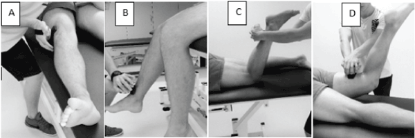
Clinical evaluation: An experienced physiotherapist performed a clinical investigation comprising measures of muscle strength, joint range of motion and functional movement performance. A handheld dynamometer (HHD) (microFET 2, Hoggan, Salt Lake City, US) was used to evaluate peak torque of different hip and knee muscles [21,22]. (Figure 1) illustrates the evaluation of the muscle force using the HHD. For the evaluation of the gluteus medius muscle, patients were positioned supine with the hip joint in a neutral anatomic position in both the frontal and sagittal plane. The investigator applied the HHD to the lateral side of the femur, 5cm proximally from the knee joint. For evaluation of the gluteus maximus, patients were in prone position with neutral hip and the knee 90° flexed. The investigator applied the HHD to the dorsal aspect of the distal part of the femur. For evaluation of knee extension peak torque, patients sat upright on an examination table with the knee flexed at 90°. The HHD was applied to the distal anterior aspect of the tibia. Knee flexion peak torque was assessed with patients lying in a prone position with the knee flexed at about 70°. The HHD was applied to the posterior aspect of the distal tibia. Patients were instructed to resist the force applied by the investigator, resulting in a maximal isometric muscle activation of the evaluated muscle (break test). The use of an HHD for the evaluation of peak muscle torque of the lower limb is shown to be reliable and valid [23,24].

Figure 1. Evaluation of peak torque using a handheld dynamometer for gluteus medius muscle (A), quadriceps (B), hamstrings (C) and gluteus maximus (D).
Active knee extension, knee flexion and ankle dorsiflexion mobility were assessed using a standard goniometer. Sagittal knee mobility was evaluated with patient’s supine: knee flexion was measured with the hip in 80° flexion, knee extension was measured with hips in a neutral position [25]. Ankle dorsiflexion was measured with patient’s supine and the knee in extension [26].
At last, subjects were asked to perform a squat and forward lunge (bilateral) three times, which were scored in real time by an experienced physiotherapist using a binary dichotomous scoring as described by Whatman et al. [11,27,28]. The second performance was used for analysis. The performance of both the lunge and squat was rated ‘good’ or ‘bad’ at trunk, pelvis and knee level. Both functional movements were evaluated as follows: good performance at trunk level was given if no deviation from the frontal or sagittal plane was observed. A good performance at pelvis level was given if no deviations were observed in the frontal or sagittal plane and if the pelvis didn’t move away from the midline. A good performance at knee level was given if the knee did not deviate from the sagittal plane (facing inward or outward). A bad score was given to subjects that couldn’t meet the criteria mentioned above.
Statistical analysis: Values for muscle force were corrected for body weight and body length using the equation described by Krause et al. [29].
Differences between groups concerning age, body weight, BMI, pain, functionality, psychosocial variables, muscle force and joint mobility were evaluated by means of an ANOVA test, followed by a post-hoc bonferroni test. Differences in gender and functional movement score between groups were calculated using a chi-square test. The relation between the performance score of the trunk or the pelvis on the one hand and the performance score of the knee on the other hand was evaluated using a chi-square test. An unpaired T-test was used to assess differences in muscle force, hamstring/quadriceps ratio (HQ) and joint mobility between patients who had a good or bad performance at knee level. For all statistical tests, significance level was set at 0.05. Post hoc test results are reported systematically for the convenience of the reader. All statistical tests were performed in SPSS version 20 (IBM, US).
Evaluation of pain, functionality and psychosocial variables: No differences were observed between groups for KOOS subscale values except for quality of life scores between both arthroplasty groups and controls. OKS-values did not differ between groups (Table 2).
Table 2. Pain and functionality scores.
|
Patient with UKA |
Patients with TKA |
Healthy controls |
p-valueb |
KOOS Pain |
89.7 (13.9) |
83.0 (25.4) |
96.3 (7.9) |
0.25 |
KOOS Symptoms |
89.3 (10.1) |
88.9 (9.2) |
93.3 (17.7) |
0.75 |
KOOS ADL |
88.9 (13.3) |
84.0 (19.2) |
97.2 (6.3) |
0.15 |
KOOS Sport |
67.9 (28.7) |
66.1 (31.2) |
90.0 (22.9) |
0.16 |
KOOS quality of life |
75.0 (17.3) |
82.6 (12.0) |
96.5 (8.3) |
0.011,2 |
OKS |
43.0 (3.7) |
42.2 (6.2) |
48.6 (6.3) |
0.06 |
KOOS=Knee injury and Osteoarthritis Outcome Score, ADL=activities of daily living, OKS=Oxford Knee Score, UKA=unicondylar knee arthroplasty, TKA=total knee arthroplasty,
b ANOVA with post hoc Bonferroni tests were performed at the significance level of .05; Post hoc p-values are presented, 1 indicates significant difference between UKA and controls, 2 indicates significant difference between TKA and controls, 3 indicates significant difference between UKA and TKA.
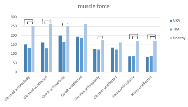
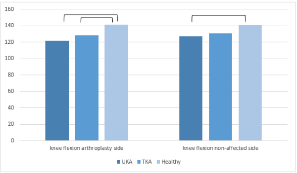
Clinical evaluation: Both knee replacement groups had lower corrected muscle peak force values compared to the control group for gluteus medius and hamstrings, at both the arthroplasty and unaffected side. Patients with TKA showed lower strength values for the quadriceps and gluteus maximus at the arthroplasty side (Figure 2). Passive ankle dorsiflexion and knee extension mobility did not differ between groups. Significant reduction of knee flexion ROM was observed at the arthroplasty side in both patient groups compared to the controls. Patients with UKA showed decreased knee flexion range of motion compared to controls at the non-affected side (Figure 3).

Figure 2. Corrected values for muscle peak torque evaluated with the HHD, significant differences between groups

Figure 3. Joint range of motion, significant differences between groups
Differences in lunge performance were observed at trunk and knee level. As many of control subjects demonstrated a good performance according to the Whatman-criteria at both trunk and knee level, whereas many subjects with knee replacement demonstrated a bad performance (Table 3). Squat performance did not differ between groups (Table 3). Additionally, a relation could be observed between the performance of the lunge at trunk and the performance at knee level. Subjects showing good performance at trunk level also performed good at knee level, whereas those subjects with a bad performance at trunk level showed a bad performance at knee level too (Table 4). This relation could not be observed at the pelvic level.
Table 3. Percentages of subjects with a good or bad performance of the forward lunge at knee, trunk and pelvis level.
|
Lunge trunk |
Lunge pelvis |
Lunge knee |
Squat trunk |
Squat pelvis |
Squat knee |
Bad |
Good |
Bad |
Good |
Bad |
Good |
Bad |
Good |
Bad |
Good |
Bad |
Good |
UKA |
85.7 |
14.2 |
71.4 |
28.5 |
100 |
0 |
14.2 |
85.7 |
0 |
100 |
42.9 |
57.1 |
TKA |
55.6 |
44.4 |
33.3 |
66.6 |
88.9 |
11.1 |
33.3 |
66.6 |
44.4 |
55.6 |
55.6 |
44.4 |
Healthy |
22.2 |
77.8 |
22.2 |
77.8 |
33.3 |
66.6 |
22.2 |
77.8 |
22.2 |
77.8 |
44.4 |
55.6 |
Sign.c |
0.04 |
0.12 |
0.01 |
0.67 |
0.12 |
0.85 |
UKA=unicondylar knee arthroplasty; TKA=Total knee arthroplasty, sign=significance level. c Chi-square tests were performed at the significance level of .05, on performance scores at knee, pelvis and knee level for both the squat and forward lunge; Bold indicates differences between ‘Good’ or ‘Bad’ performance scores at different body levels.
Table 4. Relation between performance at knee level and good or bad execution at trunk and pelvis level.
|
Bad trunk |
Good trunk |
Bad pelvis |
Good pelvis |
Bad knee |
66.7 |
33.3 |
50.0 |
50.0 |
Good knee |
14.3 |
85.7 |
14.3 |
85.7 |
Sign.c |
0.03 |
0.13 |
Sign=significance level
c Chi-square tests were performed at the significance level of .05, on performance scores at knee, pelvis and knee level for both the squat and forward lunge; Bold indicates differences between ‘Good’ or ‘Bad’ performance scores at different body levels.
At last, significant differences in muscle force and knee range of motion were demonstrated between subjects with bad and good performance of the lunge at knee level. At the arthroplasty side, the group with a bad performance of the lunge showed decreased peak torques for the gluteus medius, quadriceps, gluteus maximus and hamstrings as well as a decreased knee flexion (Table 5).
Table 5. Muscle force and knee range of motion based on lunge performance at knee level at the operated or non-dominant side.
|
Bad performance at knee level (n=18) |
Good performance at knee level (n=7) |
p-valued |
UKA/TKA/Healthy |
7/8/3 |
0/1/6 |
|
Male/Female |
9/9 |
2/5 |
0.33 |
Peak torque gluteus medius (N) |
151.6 (59.2) |
258.7 (56.1) |
0.00 |
Peak torque quadriceps (N) |
194.1 (56.1) |
264.6 (71.5) |
0.07 |
Peak torque gluteus maximus (N) |
128.4 (38.4) |
186.7 (40.2) |
0.01 |
Peak torque hamstrings (N) |
97.6 (44.6) |
162.7 (51.3) |
0.01 |
ROM knee flexion (N) |
127.2 (8.0) |
140.1 (12.3) |
0.01 |
ROM knee extension (N) |
178.9 (2.2) |
179.4 (1.0) |
0.54 |
H/Q ratio |
0.612 |
0.63 |
0.85 |
UKA=unicondylar knee arthroplasty; TKA=Total knee arthroplasty; N=Newton; H/Q ratio=hamstrings/quadriceps ratio.
d Independent t tests were performed at the significance level of .05; Bold indicates significant differences between subjects with a good or bad performance at knee level.
The main finding of the present study is that patients with knee replacement demonstrate a bad performance of the forward lunge one year after knee surgery compared to controls. This study could not demonstrate a difference in FL performance between patients with a UKA and TKA. However, almost all knee replacement patients showed a bad performance at knee level, with deviations in the frontal plane. These frontal plane deviations were related to reduced peak torque of the hamstrings, gluteus medius, gluteus maximus, quadriceps and a reduced knee flexion range of motion of the involved leg.
Findings regarding the relation between hamstrings and quadriceps muscle strength are in accordance with previous research, as it has been demonstrated that sufficient strength of these muscles is essential for a good execution of the forward lunge [30]. Noteworthy is that in this study the H/Q ratio did not significantly differ between patients who had a ‘good’ or ‘bad’ performance of the forward lunge. The strength of the individual muscles might be more important than their ratio for the execution of this task. Yet, one should take into account that in the current sample a, statistically not significant (p=), reduction in quadriceps strength could also be observed among subjects who performed the FL poorly, this could have affected the resulting H/Q ratio. Previous authors have demonstrated that both the quadriceps and hamstrings have a major role in the performance of the forward lunge [13]. Nevertheless, in order to confirm this statement more research is needed which focusses on the relation between muscle strength, muscle activation patterns and forward lunge performance in this particular population. The gluteus medius also is involved in the frontal plane control of the pelvis and lower limb during loaded activities [31,32]. A decrease in its peak torque could also be a contributing factor to the frontal plane deviations that were observed at knee level in our population [32]. Finally, Alkjaer et al. reported that the gluteus maximus, a prime stabilizer of the pelvis and trunk, has an important stabilizing function of the knee during a forward lunge [16]. Interestingly, a bad performance at knee level could also be associated with a poor execution at trunk (66.7%) and pelvis (50%) level, suggesting that the forward lunge is not merely a task that challenges the knee joint, but rather the entire biomechanical chain. Moreover, trunk position is known to influence the kinetics, kinematics as well as the resulting muscular activity patterns of muscles surrounding the knee joint [33].
The performance of the squat did not differ between subjects with a knee replacement and the age matched controls. Patients with TKA and UKA were able to perform a squat in a satisfactory way at trunk, pelvis and knee level. This could indicate that the squat is a less challenging task for knee replacement patients compared to the forward lunge. However, a recent study, which analyzed the squat movement using a 3D motion capture system, revealed significant differences in functional performance between patients with knee replacement and controls. For instance, a significant difference of 1.5s was found between TKA and controls for the duration of the descending part of the squat, or, a few mm of displacement in center of pressure mean distance has also been reported [17]. These differences can hardly be identified by visual observation alone and require more sophisticated equipment which is not readily available in daily clinical practice.
Results of the current study may shed some light on the use of functional movements during rehabilitation of knee replacement patients. At first, patients with significantly less muscle force at the gluteus medius, quadriceps and hamstrings demonstrated a worse performance of the forward lunge (Table 5). Second, the relation between the performance at trunk and knee level suggests that patients might benefit from trunk (control) exercises during rehabilitation following knee replacement. Third, the current study demonstrates reduced muscle strength at the non-affected side, suggesting that strengthening both legs is indicated. At last, rehabilitation schemes should also focus on hip extensors and hip abductors, as they play an important role in the optimal alignment of the leg during gait and functional movements such as the forward lunge and are shown to be not fully recovered one year after knee replacement surgery. In the light of these results, we believe that knee replacement patients would benefit from a rehabilitation protocol which incorporates the forward lunge exercise in addition to the squat.
This study has also some shortcomings. Despite a good reliability and validity of performance-based assessment of the forward lunge and the squat [11,27], results are dependent of the visual evaluation of the researcher. A visual rating at multiple body levels (knee, pelvis, trunk) requires a high level of concentration and accuracy from the investigator. The small sample size makes it hard to generalize the results of this study. However, the obtained results seem promising for future analysis in larger sample sizes. Last, muscle force evaluation by means of a handheld dynamometer requires sufficient counter force from the investigator in order to fully evaluate a subject’s strength, especially when assessing large muscle groups [21]. Nevertheless, the use of a handheld dynamometer in the evaluation of muscle force was found to be valid and reliable in previous studies [34-37].
This study revealed that the forward lunge is a challenging task for knee replacement patients, and more specifically for those who have reduced muscle strength at both hip and knee stabilizers (e.g. gluteus medius, quadriceps and hamstrings). These results suggest that rehabilitation should not merely focus on knee joint stability, but rather address the entire biomechanical chain. In addition, in the performance of the forward lunge, the H/Q ratio appears to be less important than the individual muscle strength of the hamstrings and quadriceps. No differences were found for squat performance between controls and knee replacement patients, which could indicate that this task is less challenging than the forward lunge for this population. Moreover, it is our opinion that knee arthroplasty patients would benefit from a rehabilitation protocol which incorporates the forward lunge as this will activate different muscle groups which are recruited whilst performing other functional movements such as the squat. Future studies should further investigate the impact of underlying kinematics, kinetics and muscle recruitment patterns on the performance of the forward lunge and squat.
This research was supported by European Regional Development Fund, project: grant number 1047.
- Koskinen E, Eskelinen A, Paavolainen P, Pulkkinen P, Remes V (2008) Comparison of survival and costeffectiveness between unicondylar arthroplasty and total knee arthroplasty in patients with primary osteoarthritis: A followup study of 50,493 knee replacements from the Finnish Arthroplasty Register. Acta Orthop 79: 499-507. [Crossref]
- Willems T (2017) Orthopride Belgian Hip and Knee Arthroplasty Registry Annual Report 2016. Belgium: BVOT.
- Pabinger C, Lothaller H, Geissler A (2015) Utilization rates of knee-arthroplasty in OECD countries. Osteoarthritis Cartilage 23: 1664-1673. [Crossref]
- Longo UG, Loppini M, Trovato U, Rizzello G, Maffulli N, et al. (2015) No difference between unicompartmental versus total knee arthroplasty for the management of medial osteoarthtritis of the knee in the same patient: a systematic review and pooling data analysis. Br Med Bull 114: 65-73. [Crossref]
- Ranawat CS (2002) History of total knee replacement. J South Orthop Assoc 11: 218-226. [Crossref]
- Bonnin M, Amendola A, Bellemans J, MacDonald S, Menetrey (2012) The Knee Joint: Surgical Techniques and Strategies. Springer Paris: Imprint Springer, Paris.
- Fabre-Aubrespy M, Ollivier M, Pesenti S, Parratte S, Argenson JN (2016) Unicompartmental Knee Arthroplasty in Patients Older Than 75 Results in Better Clinical Outcomes and Similar Survivorship Compared to Total Knee Arthroplasty. A Matched Controlled Study. J Arthroplasty 31: 2668-2671. [Crossref]
- Lyons MC, MacDonald SJ, Somerville LE, Naudie DD, McCalden RW (2012) Unicompartmental versus total knee arthroplasty database analysis: is there a winner? Clin Orthop Relat Res 470: 84-90. [Crossref]
- Komnik I, Weiss S, Fantini Pagani CH, Potthast W (2015) Motion analysis of patients after knee arthroplasty during activities of daily living- A systematic review. Gait Posture 41: 370-377. [Crossref]
- Engh GA, Parks NL, Whitney CE (2014) A Prospective Randomized Study of Bicompartmental vs. Total Knee Arthroplasty with Functional Testing and Short-Term Outcome. J Arthroplasty 29: 1790-1794. [Crossref]
- Whatman C, Hume P, Hing W (2013) The reliability and validity of physiotherapist visual rating of dynamic pelvis and knee alignment in young athletes. Phys Ther Sport 14: 168-174. [Crossref]
- Riemann B, Congleton A, Ward R, Davies GJ (2013) Biomechanical comparison of forward and lateral lunges at varying step lengths. J Sports Med Phys Fitness 53: 130-138. [Crossref]
- Jonhagen S, Halvorsen K, Benoit DL (2009) Muscle activation and length changes during two lunge exercises: implications for rehabilitation. Scand J Med Sci Sports 19: 561-568. [Crossref]
- Thorlund BJ, Damgaard MJ, Roos ME, Aagaard P (2012) Neuromuscular Function during a Forward Lunge in Meniscectomized Patients. Med Sci Sport Exer 44: 1358-1365.
- McClelland JA, Feller JA, Menz HB, Webster KE (2017) Patients with total knee arthroplasty do not use all of their available range of knee flexion during functional activities. Clin Biomech (Bristol, Avon) 43: 74-78. [Crossref]
- Alkjaer T, Wieland MR, Andersen MS, Simonsen EB, Rasmussen J (2012) Computational modeling of a forward lunge: towards a better understanding of the function of the cruciate ligaments. J of Anatomy 221: 590-597.
- Verdini F, Zara C, Leo T, Mengarelli A, Cardarelli S, et al. (2017) Assessment of patient functional performance in different knee arthroplasty designs during unconstrained squat. Muscles Ligaments Tendons J 7: 514-523. [Crossref]
- Roos EM, Roos HP, Lohmander LS, Ekdahl C, Beynnon BD (1998) Knee Injury and Osteoarthritis Outcome Score (KOOS)--development of a self-administered outcome measure. J Orthop Sports Phys Ther 28: 88-96. [Crossref]
- Dawson J, Fitzpatrick R, Murray D, Carr A (1998) Questionnaire on the perceptions of patients about total knee replacement. J Bone Joint Surg Br 80: 63-69. [Crossref]
- Miller RP, Kori SH, Todd DD (1991) The Tampa Scale: a Measure of Kinisophobia. The Clin J of Pain 7: 51.
- Mentiplay B, Perraton L, Bower KJ, Adair B, Pua YH, et al. (2015) Assessment of Lower Limb Muscle Strength and Power Using Hand- Held and Fixed Dynamometry: A Reliability and Validity Study. PLoS One 10: e0140822. [Crossref]
- Stark T, Walker B, Phillips JK, Fejer R, Beck R (2011) Hand-held dynamometry correlation with the gold standard isokinetic dynamometry: a systematic review. PMR 3: 472-479. [Crossref]
- Whiteley R, Jacobsen P, Prior S, Skazalski C, Otten R, et al. (2012) Correlation of isokinetic and novel hand-held dynamometry measures of knee flexion and extension strength testing. J Sci Med Sport 15: 444-450. [Crossref]
- Kwoh CK, Petrick MA, Munin MC (1997) Inter-rater reliability for function and strength measurements in the acute care hospital after elective hip and knee arthroplasty. Arthritis Care Res 10: 128-134. [Crossref]
- Brosseau L, Balmer S, Tousignant M, O'Sullivan JP, Goudreault C, et al. (2001) Intra- and intertester reliability and criterion validity of the parallelogram and universal goniometers for measuring maximum active knee flexion and extension of patients with knee restrictions. Arch Phys Med Rehabil 82: 396-402.
- Donnery J, Spencer RB (1988) The Biplane Goniometer. A new device for measurement of ankle dorsiflexion. J Am Podiatr Med Assoc 78: 348-351. [Crossref]
- Whatman C, Hing W, Hume P (2012) Physiotherapist agreement when visually rating movement quality during lower extremity functional screening tests. Phys Ther Sport 13: 87-96. [Crossref]
- Whatman C, Hume P, Hing W (2013) Kinematics during lower extremity functional screening tests in young athletes - Are they reliable and valid? Phys Ther Sport 14: 87-93. [Crossref]
- Krause DA, Schlagel SJ, Stember BM, Zoetewey JE, Hollman JH (2007) Influence of Lever Arm and Stabilization on Measures of Hip Abduction and Adduction Torque Obtained by Hand- Held Dynamometry. Arch Phys Med Rehabil 88: 37-42. [Crossref]
- Thorlund JB, Damgaard J, Roos EM, Aagaard P (2012) Neuromuscular function during a forward lunge in meniscectomized patients. Med Sci Sports Exerc 44: 1358-1365. [Crossref]
- Kim D, Unger J, Lanovaz JL, Oates AR (2016) The Relationship of Anticipatory Gluteus Medius Activity to Pelvic and Knee Stability in the Transition to Single-Leg Stance. PMR 8: 138-144. [Crossref]
- Lubahn AJ, Kernozek TW, Tyson TL, Merkitch KW, Reutemann P, et al. (2011) Hip muscle activation and knee frontal plane motion during weight bearing therapeutic exercises. Int J Sports Phys Ther 6: 92-103. [Crossref]
- Farrokhi S, Pollard CD, Souza RB, Chen YJ, Reischl S, et al. (2008) Trunk position influences the kinematics, kinetics, and muscle activity of the lead lower extremity during the forward lunge exercise. J Orthop Sports Phys Ther 38: 403-409. [Crossref]
- Bandinelli S, Benvenuti E, Del Lungo I, Baccini M, Benvenuti F, et al. (1999) Measuring muscular strength of the lower limbs by hand-held dynamometer: a standard protocol. Aging (Milano) 11: 287-293. [Crossref]
- Bohannon RW (1990) Hand-held compared with isokinetic dynamometry for measurement of static knee extension torque (parallel reliability of dynamometers). Clin Phys Physiol Meas 11: 217-222. [Crossref]
- Bohannon RW (1986) Test-retest reliability of hand-held dynamometry during a single session of strength assessment. Phys Ther 66: 206-209. [Crossref]
- Bohannon RW, Andrews AW (1987) Interrater reliability of hand-held dynamometry. Phys Ther 67: 931-933. [Crossref]


