Abstract
Non-Hodgkin Lymphoma (NHL) is one of the rare malign diseases that can affect the oral cavity. In extranodal manifestations cranial bone could be affected by a massive destruction of bone tissue and invasion of the surrounding soft tissues. 3D radiology can help identifying suspicious lesions at an early stage and can support immunohistochemical examinations. Identifying clinically or radiologically these lesions and diagnosing them means saving patients’ lives. A clinical case of extranodal oral NHL manifestation, with remission at 5 years of follow-up, identified due to a Cone Beam Computed Tomography (CBCT) examination will be presented.
key words
cone beam computed tomography, mandible lymphoma, non hodgkin’s lymphoma
Introduction
Over the years, tumors have undergone a noticeable increase, probably also due to the increase in diagnostic possibilities [1]. OSCC represents more than 90% of malignant lesions of the oral cavity [2,3]. In minor percentage, it was found other non-epithelial neoplastic lesions [4,5]. These included lymphomas, tumor metastases, sarcomas and other rare forms of neoplastic Given the rarity of these injuries it was important to recognize a correct and early diagnosis. They were often in differential diagnosis with non-neoplastic lesions and if not recognized they could interfere with diagnosis and treatment [4,5]. Among the rare tumors it can be found the malignant lymphomas in two different types: Hodgkin Lymphoma (HL) and Non-Hodgkin Lymphoma (NHL). These can manifest in several clinical forms and with different histological characteristics [6]. NHL may have different manifestations within lymph nodes or extra-nodal onset [7]. NHL forms included B-cell, T-cell and more rarely NK-cell lymphomas [6,7]. Up to 75% of NHL cases occur in the head and neck area. In these locations, NHL can arise with almost identical distribution of lymph node sites (55%) and extra nodal sites (45%) [8]. In extranodal manifestations, the first site of systemic onset is the gastro intestinal tract. Secondarily, the mainly district affected is the head-neck district which specifically can be hit in different areas such as: mandible, gingiva, hard palate, tongue, nose and paranasal sinuses [9]. Generally, oral NHL could present painful, swelling and ulcerate mucosa that could affect bone volumes, as well [10]. Taking into consideration the latter, in daily clinical practices it is possible to intercept extranodal NHL of the oral cavity with very variable clinical manifestations.
The prognosis of NHL is extremely variable, depending on the grading and aggressiveness of the lesion. Invasion of other organs or delayed diagnosis may change the 5-year prognosis ranging from 83% to 49% [11,12]. Therefore, appropriate chemotherapy and radiotherapy treatment is essential to improve survival rate. Knowing and recognizing clinical and radiological manifestations at an early stage could help in the diagnosis and improve patients’ prognosis. The aim of this case report is to present a case report of extranodal oral manifestation of NHL with a 5-year follow-up to better understand how to intercept these lesions in the early phases
Case presentation
A 36-year-old man in good general health, smoker, comes at the dental clinic of the Department of Medical Oral and Biotechnological Sciences, for a check-up visit on May 2012. No intra or extra oral pathologies were reported. Panoramic radiograph revealed a radiolucent apical lesion in correspondence of teeth 41, 31, 32, 33 (Figure 1). No positivity on these dental elements in terms of mobility, periodontal probing and pain during percussion test was found. The teeth 32 and 33 were negative at the vitality tests so it was decided to perform root canal treatments. Approximately 6 months later, a periapical control radiograph was performed showing a healing lesion in correspondence of these two teeth (Figure 2). Occasionally, during a routine control visit, a similar radiolucent lesion was found interesting the elements 34, 35, 36 absent at the initial radiographs (Figure 3). Once more, vitality test was performed and the dental elements 34 and 35 appeared necrotic. They also had a high degree of mobility (absent six months earlier). A Cone Beam Computed Tomography (CBCT) examination was performed to better investigate the lesion extension. CBCT showed the presence of two different radiolucent lesions of the mandible close to teeth 34, 35, 36 and 42, 43 (Figure 4). In both cases there was an involvement of the buccal cortex which had also shifted the root of teeth 43 and 35 as shown on Figure 4. Only in the right side there was a slight swelling of the vestibule, completely absent in the left side (Figure 5). Teeth 42, 43 were also vital, meanwhile teeth 34, 35, had loose the pulp vitality whilst the 36 was treated previously. Excluding the periapical inflammatory origin, it was decided to perform a biopsy of the lesions. It was decided to proceed with a surgical access and biopsy of the two lesions to better understand the nature of them. The collected material clinically appeared to be fibrous and poorly organized. In the right side, there was an involvement of the soft tissues overlying the missing cortical bone. The overlying gingiva was also collected. The dental elements did not appear to be macroscopic affected by the lesion. The wound was sutured, and the sutures removed after 7 days. The samples were sent to the pathologist. Histological examination revealed disorganized bone tissue with neoplastic infiltration elements. Sample showed atypical lymphoid cells with medium and large size with irregular-shaped nucleus. Also, there were blast cells that dissected the muscular bundles into soft tissue biopsy. Immunohistochemical investigation was also performed, revealing a widespread positivity of the CD20, CD3 and BCL-6, neoplastic population markers. Moreover, negativity for cytokeratin (Pan) was shown. Meanwhile, a high positivity (60-70%) for Ki67 proliferation marker was observed. Based on the light microscopic and immunohistochemical findings, a large-cell B lymphocytes lymphoma with high proliferative kinetics was diagnosed.
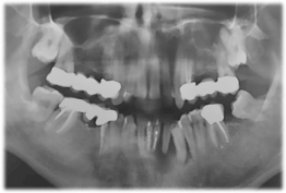
Figure 1. Orthopantomography image showing the radio transparency in correspondence of the teeth at the first appointment
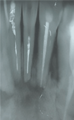
Figure 2. Intraoral periapical radiography showing the healing of the initial lesion after 6 months of treatment
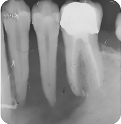
Figure 3. New radio transparency lesion interesting the teeth 34, 35 and 36 after 6 months of follow up.
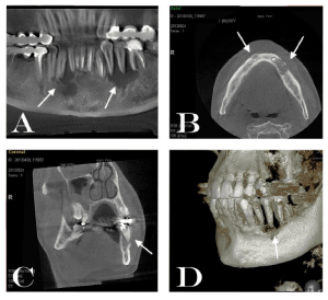
Figure 4. CBCT images showing at: A) the extension of the lesions through simil panorex images; B) coronal CBCT images indicating the erosion of the vestibular cortical plate; C) the axial image showing the displacement of the root of 35 as indicated by the arrow; D) 3D reconstruction of the lesion in the area of 34, 35 and 36.
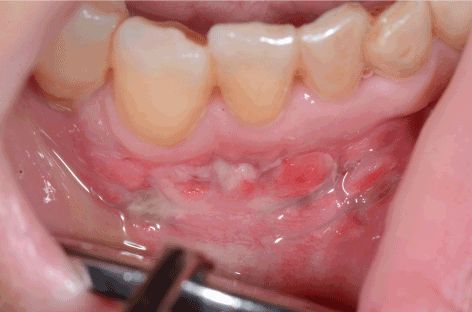
Figure 5. Clinical image showing the involvement of the soft tissues surrounding the teeth 42 and 43
PET-CT examination was also performed (Figure 6). The latter demonstrated a metabolic activity in the mandibular body and in the surrounding soft tissues. No activity has been reported in other different areas of the body. Following the confirmation of the first diagnosis, the therapy was started. R-Chop chemotherapy and 20 sessions of radiotherapy of the jaw were immediately performed. Over the years, the patient has been kept under observation in the reference hematology center and under dental observation in our clinic. Five years after the diagnosis of the pathology, clinical examination, radiological exam and blood test appeared to be normal and the patient was declared healed with complete remission of the disease.
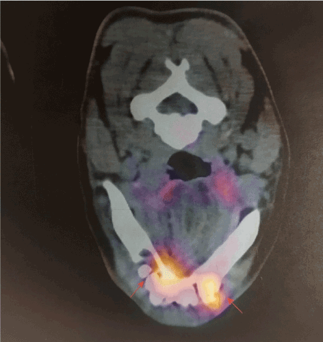
Figure 6. Pet tc demonstrating the positiveness of the lesions on the lower jaw
Discussion and conclusion
In the present case report an extra nodal NL with oral manifestations was reported. The origin and the only site of manifestation of the tumor was the mandible. The growth occurred within the mandible with minimal involvement of the soft tissues overlying the right side of jaw. Meanwhile, no soft tissue involvement was observed on left side. Although, the extra nodal manifestations of NHL are more frequent than HL, the primary oral manifestation were quite rare and poorly described [11]. In any case, the most described typology is B-cell lymphoma, as in our case [11,12]. The NHL localization in the head-neck district was reported to be 25% with a high manifestation especially on tonsils and oropharynx [13]. Therefore, the case described here represented a rare localization with hard clinical interpretation. The concomitance with inflammatory pathologies, could make difficult the diagnostic process. Thus, differential diagnosis became a key moment in the diagnostic process of specific pathologies that could simulate inflammatory manifestations both in radiological and clinical aspect. It also has been demonstrated that the immunohistochemical investigation became a very important moment in the differential diagnosis and in the staging of this kind of pathology [14]. Immunohistochemical techniques to date can also be used in the diagnostic framework of inflammatory diseases [15,16]. Together with traditional immunohistochemical techniques, alternative methods and specific markers for the classification of the pathology was used more frequently [17,18]. Specifically, in the histological findings cellular and nuclear anomalies can be found, mixed with tissue with ischemic necrosis and vascular destruction [14]. In any case the immunohistochemical investigation of CD20, CD3, Ki-67 proliferation marker, BCL-6 and BLC-2, intracellular T-cell antigen (TIA-1) may suggest a high proliferative and cytotoxic activity, suggesting aggressiveness and malignant of the lesion [14]. In the present case it is important to remember how the patient appeared completely asymptomatic. Moreover, at least two-thirds of NHL cases present a persistent enlargement of the lymph nodes for more than two weeks [19]. Systemic symptoms such as high fever, nonspecific weight loss, general pain and illness was also associated. In our, early intercepted, case no systemic symptoms were associated [20]. Clinicians, as in the case described, must pay attention to all the oral manifestations of NHL in order to achieve an early diagnosis and treatment planning. It was important to recognize the radiographic signs sometimes present as roots shift, mobility and massive erosion of the cortical bone. Even in the absence of radiographic signs, it is necessary to intercept prematurely painless, soft and ulcerated swelling. Attention to these evaluations can greatly improve the prognosis which is otherwise unfavorable [21].
An early diagnosis, carried out thanks to a careful clinical examination and radiological findings, allowed to perform an early treatment plan and save the patient's life. The 5-year survival rate, changes from 73% in HL to 65% in extranodal head and neck NHL sites. The prognosis decreases due to a possible delayed diagnosis that could lead to a greater spread of the disease [12,22].
Despite the manifestation in the head and neck of NHL which is the second for frequency after the gastrointestinal tract, it is considered a very rare disease [11]. Therefore, an appropriate description of radiological images in the literature was not very exhaustive [21]. It has been described that mandibular localization can lead to an ill-defined bone lesion on conventional panoramic radiographs [21]. In the early manifestations, as in our case, the radiological images were comparable to inflammatory periapical pathologies affecting the surrounding teeth. At the CBCT exam, however, there were signs suggesting malignancy: occlusal displacement of the teeth, loss of the lamina dura, enlargement of the periodontal space, destruction of the cortical bone [23]. This last sign was more pronounced on the side associated with a lesion of the overlying soft tissue. Different studies associated the erosion of the cortical bone to NHL even in long bones [24,25]. On the contrary, an exclusively gingival localization was associated, thanks to the CBCT, with micro-erosion of the underlying cortical bone [26].
This suggests the importance of the CBCT exam in early diagnosis and in the possibility of identifying initial bone involvement. Therefore, the use of CBCT, respect to the panoramic radiography, was necessary in the diagnosis of initial and small lesions. In conclusion, we recommend the use of CBCT associated with other diagnostic methods to evaluate not only bone lesions but also bone micro-infiltrations in lesions affecting soft tissues. Differential diagnosis with other diseases that may affect the jaw as multiple myeloma, osteosarcoma, osteomyelitis or bone metastasis, can be difficult [24,25]. Surely, the histological and immunohistochemical investigations are mandatory to make a diagnosis, but the CBCT exam could help to investigate radiolucent lesions that were not clearly classifiable, greatly improving the diagnostic possibilities and therefore the prognosis of the pathology.
References
- Rivera C (2015) Essentials of oral cancer. Int J Clin Exp Pathol 8: 11884-11894. [Crossref]
- Choi S, Myers JN (2008) Molecular pathogenesis of oral squamous cell carcinoma: implications for therapy. J Dent Res 87: 14-32. [Crossref]
- Weijers M, Leemans CR, Aartman IH, Karagozoglu KH, van der Waal I (2011) Oral cancer trends in a single head-and-neck cancer center in the Netherlands; decline in T-stage at the time of admission. Med Oral Patol Oral Cir Bucal 16: e914-918. [Crossref]
- Bagan J, Sarrion G, Jimenez Y (2010) Oral cancer: clinical features. Oral Oncol 46: 414-417. [Crossref]
- Scully C, Bagan J (2009) Oral squamous cell carcinoma overview. Oral Oncol 45: 301-308. [Crossref]
- Müller AM, Ihorst G, Mertelsmann R, Engelhardt M (2005) Epidemiology of non-Hodgkin's lymphoma (NHL): trends, geographic distribution, and etiology. Ann Hematol 84: 1-12. [Crossref]
- http://publications.iarc.fr/Book-And-Report-Series/Who-Iarc-Classification-Of-Tumours/Who-Classification-Of-Tumours-Of-Haematopoietic-And-Lymphoid-Tissues-2017
- Iguchi H, Wada T, Matsushita N, Oishi M, Yamane H (2012) Anatomic distribution of hematolymphoid malignancies in the head and neck: 7 years of experience with 122 patients in a single institution. Acta Otolaryngol 132: 1224-1231. [Crossref]
2021 Copyright OAT. All rights reserv
- Ratech H, Burke JS, Blayney DW, Sheibani K, Rappaport H (1989) A clinicopathologic study of malignant lymphomas of the nose, paranasal sinuses, and hard palate, including cases of lethal midline granuloma. Cancer 64: 2525–2531. [Crossref]
- Vega F, Lin P, Medeiros LJ (2005) Extranodal lymphomas of the head and neck. Ann Diagn Pathol 9: 340-350. [Crossref]
- Zapater E, Bagán JV, Carbonell F, Basterra J (2010) Malignant lymphoma of the head and neck. Oral Dis 16: 119-128. [Crossref]
- Economopoulos T, Asprou N, Stathakis N, Fountzilas G, Pavlidis N, et al. (1992) Primary extranodal non-Hodgkin's lymphoma of the head and neck. Oncology 49: 484-488. [Crossref]
- Epstein JB, Epstein JD, Le ND, Gorsky M (2001) Characteristics of oral and paraoral malignant lymphoma: A population-based review of 361 cases. Oral Surg Oral Med Oral Pathol Oral Radiol Endod 92: 519. [Crossref]
- Tan LH (2009) A practical approach to the understanding and diagnosis of lymphoma: an assessment of the WHO classification based on immunoarchitecture and immuno-ontogenic principles. Pathology 41: e305-e326. [Crossref]
- Álvares PR, Arruda JAA, Silva LPD, Nascimento GJFD, Silveira MFD, et al. (2017) Immunohistochemical expression of TGF-ß1 and MMP-9 in periapical lesions. Braz Oral Res 31: e51. [Crossref]
- D'addazio G, Artese L, Piccirilli M, Perfetti G (2014) Role of matrix metalloproteinases in radicular cysts and periapical granulomas. Minerva Stomatol 63: 411-20.
- D'Addazio G, Artese L, Traini T, Rubini C, Caputi S, et al. (2018) Immunohistochemical study of osteopontin in oral squamous cell carcinoma allied to fractal dimension. J Biol Regul Homeost Agents 32: 1033-1038. [Crossref]
- Mincione G, Di Nicola M, Di Marcantonio MC, Muraro R, Piattelli A, et al. (2015) Nuclear fractal dimension in oral squamous cell carcinoma: a novel method for the evaluation of grading, staging, and survival. J Oral Pathol Med 44: 680-684. [Crossref]
- Mawardi H, Cutler C, Treister N (2009) Medical management update: Non-Hodgkin lymphoma. Oral Surg Oral Med Oral Pathol Oral Radiol Endod 107: e19-e33. [Crossref]
- Van der Waal RI, Huijgens PC, Van der Valk P, van der Waal I (2005) Characteristics of 40 primary extranodal non-Hodgkin lymphomas of the oral cavity in perspective of the new WHO classification and the International prognostic index. Int J Oral Maxillofac Surg 34: 391-395. [Crossref]
- Imaizumi A, Kuribayashi A, Watanabe H, Ohbayashi N, Nakamura S, et al. (2012) Non-Hodgkin lymphoma involving the mandible: imaging findings. Oral Surg Oral Med Oral Pathol Oral Radiol 113: e33-e39. [Crossref]
- Urquhart A, Berg R (2001) Hodgkin's and non-Hodgkin's lymphoma of the head and neck. Laryngoscope 111: 1565-1569. [Crossref]
- Wood RE (2009) Malignant diseases of the jaws. (6th edn). St. Louis: Mosby: 405-427.
- Mulligan ME, McRae GA, Murphey MD (1999) Imaging features of primary lymphoma of bone. AJR Am J Roentgenol 173: 1691-1697. [Crossref]
- Krishnan A, Shirkhoda A, Tehranzadeh J, Armin AR, Irwin R, et al. (2003) Primary bone lymphoma: radiographic-MR imaging correlation. Radiographics 23: 1371-1383. [Crossref]
- Yasumoto M, Shibuya H, Fukuda H, Takeda M, Mukai T, et al. (1998) Malignant lymphoma of the gingiva: MR evaluation. AJNR Am J Neuroradiol 19: 723-727. [Crossref]


