Three-dimensional gait analysis in 6 patients with spastic type of bilateral cerebral palsy was performed in this study. We evaluated the extension angle of the hip joint in the late stance phase, extension moment of the hip joint in the middle stance phase, and plantar flexion moment of the ankle joint in the late stance phase. The characteristics of walking after surgery were poor extension of the hip joint, reduction of extension moment of the hip joint compared to that during steady walking. In 3 of the 6 patients, the maximal extension moment was increased by fixation of the orthosis, though extension of the hip joint was not obtained even by fixation of the orthosis.
In 4 of the 6 patients, the plantar flexion moment of the ankle joint was increased by fixation of the orthosis, though reduction of plantar flexion moment in the middle stance phase was observed compared to that of steady walk. Objective evaluation of changes in the walking condition before and after application of orthoses was possible by this method.
three-dimension gait analysis, cerebral palsy, moment, orthoses, myotony
Cerebral paralysis is abnormalities in intelligence or physical capabilities caused by damage to the brain during the perinatal period. In the treatment of dyskinesia caused by cerebral paralysis, hypertonia and articular contracture, which induce abnormal walking, are important. Even though hypertonia is not detected in the decubitus, abnormal hypertonia is often observed in the standing position or during walking, for which antigravitational action is required. Therefore, evaluation in the standing position and during walking is necessary.
Bobath [1] reported that since imbalance is likely to occur in children with spastic type of bilateral cerebral palsy, walking was stabilized by posturing with which balance of the body is relatively good (bending the body trunk forward with hip flexion). Davids [2] analyzed walking of children with and without cerebral paralysis, and reported that propulsion of walking was mainly generated by the hip and ankle joints.
Smooth walking with sufficient extension of the hip joint is a factor affecting the gait, and propulsion generated mainly by extension of the hip joint and plantar flexion moment of the ankle joint is another factor.
In this study, we evaluated the following 3 items in children with cerebral paralysis who had undergone surgical treatment using a three-dimensional gait analyzer: the extension angle of the hip joint in the late stance phase, extension moment of the hip joint in the middle stance phase, and plantar flexion moment of the ankle joint in the late stance phase. Effects of orthoses were also evaluated by analyzing the same items.
The subjects were 6 patients with spastic type of bilateral cerebral palsy, consisting of 3 boys and 3 girls, with a mean age of 8.8 years (7-12 years). Four patients walked without assistance, and 2 walked with a stick. This evaluation was performed 12.8 months on average after surgery (7-23 months). The orthoses used were an ankle foot orthosis (AFO) in 3 patients and a foot orthosis (FO) in 3 patients. Postoperatively, barefoot walking was first evaluated, and then, walking with the orthosis was analyzed. All subjects were children with mild mental retardation who were cooperative in analysis of walking (Table 1). The figures shown in the Results section show data of patient 3 (Table 1). The steady walking shown in the figures was obtained by converting the graph of normal adult walking using the body weight of patient 3.
Table 1. Subject data.
Case |
sex |
age |
gait |
operation |
post op term |
orthoses |
1 |
M |
7y1mo |
W |
foot (VP) |
8mo |
AFO |
2 |
F |
12y4mo |
W |
knee (Gr Sm St), foot(VP) |
7mo |
AFO |
3 |
F |
10y8mo |
S |
hip (Gr St Sm RF AL), DVO |
11mo |
FO |
4 |
M |
12y5mo |
W |
hip (RF AL) knee (Gr Sm St), foot(Baker) |
23mo |
FO |
5 |
F |
12y6mo |
S |
hip (Gr AL RF Ps) knee (Gr St Sm), foot (VP) |
9mo |
FO |
6 |
M |
10y9mo |
W |
foot (Baker+FHL transfer) |
9mo |
AFO |
W: walk without assistance; S: gait with a stick; VP: Valpius; Gr: gracilis muscle; St: semitendinosus muscle; Sm: semimenbranosus muscle; Ps: major psoas muscle; RF: rectus femoral muscle; AL: adductor long muscle; FHL: flexor hallucis long muscle; DVO: deversion osteotomy
A three-dimensional gait analyzer (VICON 140, Oxford Metrics Inc.) was used. A ground reaction meter (Kistlar Inc.) was fixed to the analyzer, and the ground reaction was measured by synchronizing the apparatus with the movement of the patient. The movement of the patient was measured using 10 reflective markers attached to the body surface. The reflective markers were covered with microglass beads, and incident light was reflected in the same direction as the incidence. In this system, a light source was fixed to the camera, and the movement of the patient was recorded by recognizing the light source by the camera. Data were analyzed using dedicated software, and graphically represented using spreadsheet software.
Preoperative walking patterns in the children with spastic type of bilateral cerebral palsy
Figure 1 shows the graph of the preoperative articular moment in patient 3. In the figure, a cooperative pattern simultaneously showing extension moments of the hip and ankle joints, and plantar flexion moment of the ankle joint is observed. (In the following figures, angles in the direction of flexion of the hip joint and dorsiflexion of the ankle joint were taken as positive, and moments in the direction of extension of the hip joint and plantar flexion of the ankle joint were also taken as positive.)
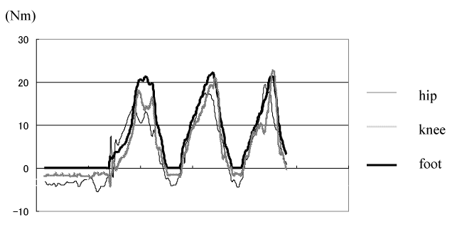
Figure 1. Pre-operation moment of spastic CP patient on barefoot walking (case 3). Hip, knee and ankle moment show the same flexion and extension moment pattern at the same time on foot.
Characteristics of postoperative walking patterns in the children with spastic type of bilateral cerebral palsy and changes in the walking patterns caused by fixation of the orthosis
Hip joint in the late stance phase and the extension angle in the swing phase: Of the 6 patients with spastic type of bilateral cerebral palsy, walking with bended legs, in which no extension of the hip joint occurs during the period between the foot flat (FF) and toe off (TO), was postoperatively observed in 3 patients (Figure 2). In the 3 patients, extension of the hip joint was not obtained even by fixation of the orthosis. The changes in the maximal extension angle ranged from 0.2° to 5.0° although improvement or aggravation was observed in the patients (Table 2).
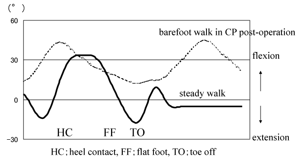
Figure 2. Hip angle on steady walk and barefoot walk in CP post-operation (case 3). No extension of the hip joint occurs during the period between the foot flat (FF) and toe off (TO), was postoperatively observed in three patients in this study.
Table 2. Change of the maximum hip flexion angle between the foot flat and toe off on gait with and without splint.
Case (gait/orthoses) |
barefoot post op |
using splint |
change |
1(W/A) |
17.0°(E ) |
11.8°(E ) |
5.2°decrease |
2(W/A) |
3.2° (E ) |
2.1°(E ) |
0.9°decrease |
3(S/F ) |
10.0° (F ) |
12.1°(F ) |
2.1°decrease |
4(W/F ) |
3.7°(F ) |
1.2°(F ) |
2.5°increase |
5(S/A) |
12.8° (F ) |
10.3°(F ) |
2.5°increase |
6(W/A) |
8.6° (E ) |
8.9°(E ) |
0.2°increase |
W: walk without assistance; S: gait with a stick; E: extension; F: flexion; A: AFO; F: FO
Extension moment of the hip joint in the middle stance phase: In the patients with spastic type of bilateral cerebral palsy, the extension of the hip joint was slightly restricted after surgery, and the extension moment of the hip joint was weak compared to that during steady walking (Figure 3). The mean maximal extension moment during barefoot walking was 27.3 Nm. In 3 of the 6 patients, the maximal extension moment was increased by fixation of the orthosis. The mean maximal extension moment during walking with the orthosis was 28.0 Nm, which was higher by 0.7 Nm than that during barefoot walking (Table 3).
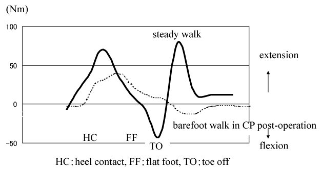
Figure 3. Hip moment on steady walk and barefoot walk in CP post-operation (case 3). The extension moment of the hip joint was weak compared to that during steady walking because of limitation in hip angle even after surgery.
Table 3. Change of the maximum hip moment between the foot flat and toe off on gait with and without splint.
Case (gait/orthoses) |
barefoot post op (Nm) |
using splint (Nm) |
change |
1(W/A) |
8.5 |
11 |
29.0% |
2(W/A) |
19.8 |
30.5 |
54.0% |
3(S/F) |
38.2 |
46.2 |
21.1% |
4(W/F) |
40 |
37.3 |
-6.7% |
5(S/A) |
31 |
12.5 |
-59.6% |
6(W/A) |
30.7 |
26.4 |
-14.0% |
average |
27.3 |
28 |
2.6% |
W: walk without assistance; S: gait with a stick; A: AFO; F: FO
Plantar flexion moment of the ankle joint in the late stance phase: The mean plantar flexion moment of the ankle joint was 55.5 Nm during barefoot walking after operation, and reduction of plantar flexion moment in the middle stance phase was observed compared to that of steady walk (Table 4, Figure 4). In 4 of the 6 patients, the plantar flexion moment of the ankle joint was increased by fixation of the orthosis, and the mean increase in the plantar flexion moment was 9.4 Nm (16.9%) (Table 4).

Figure 4. Ankle moment on steady walk and barefoot walk in CP post-operation (case 3).
Table 4. Change of the maximum ankle planter moment in the stance phase with and without splint.
Case (gait/orthoses) |
barefoot post op (Nm) |
using splint (Nm) |
change |
1(W/A) |
45.3 |
46.3 |
2.20% |
2(W/A) |
27.2 |
19.5 |
-28.30% |
3(S/F) |
19.6 |
37.6 |
91.80% |
4(W/F) |
77 |
119.4 |
55.00% |
5(S/A) |
60.8 |
49.4 |
-18.70% |
6(W/A) |
102.8 |
117.2 |
14.00% |
average |
55.5 |
64.9 |
16.9% |
W: walk without assistance; S: gait with a stick; A: AFO; F: FO
The purpose of physiotherapy and ergotherapy (rehabilitation) is to obtain suppression of myotony, enhancement of voluntary, skilled, and antigravitational movement, and sense of equilibrium, while the purpose of surgical treatment is to achieve suppression of myotony and improvement of articular contracture. We have objectively evaluated the changes in the walking condition by physiotherapy, ergotherapy, and surgery using a three-dimensional gait analyzer. Gait analysis is considered very important in determining surgical procedures and evaluating treatment effects.
Aptekar, et al. [3] examined movement patterns in children with and without disorder using photographed trajectories of small electric bulbs fixed to the head, body trunk, and 4 limbs. Simon, et al. [4] found pathologic walking and abnormal compensatory function using electromyograms, a ground reaction meter, and a 16-mm high-speed camera. Since the VICON 140 analyzer used in the present study could evaluate movement of the lower limbs by synchronizing a ground reaction meter, moment of each joint could be measured. Therefore, this method was considered more useful for the evaluation of movement in children with cerebral paralysis than conventional methods, such as the stick picture method.
Preoperatively, a posture of bended legs, which is caused by labyrinthine flexion tonus, is characteristic of children with spastic type of bilateral cerebral palsy. Walking in this posture is possible by extension and plantar flexion moments of the hip, knee, and ankle joints induced by primitive reflex, such as spontaneous extensor tension. This reflex is cooperative extension without movement of the individual joints. In this type of walking, the hip, knee, and ankle joints extend simultaneously without separated movement of these joints, which is observed during steady walking. The cooperative extension pattern was observed in the preoperative articular moment, i.e., simultaneous extension moments of the joints shown in this study.
In patients in whom suppression of articular contracture or excess myotony is insufficient by rehabilitation (physiotherapy, ergotherapy), resulting in no improvement of the walking condition, surgical treatment is often performed. To obtain suppression of myotony, improvement of articular contracture, propulsion of walking, or improvement of the walking condition, we perform surgery. In patients with a bending or adducent contracture of more than 30° observed in the hip joint by the Thomas test, detachment of muscles around the hip joint, such as incision of adductor muscles, and extension of iliopsoas muscles and rectus muscles of the thigh, was performed [5,6]. In the knee joint with a bending contracture of more than 30° in the supine position, medial hamstring muscles were partially extended [7], and detachment of the distal lacertus of gastrocnemius muscles (Baker, Vulpius method) was performed in the ankle joint with a dorsiflexion angle of less than 0° in the passive incomplete extension of the knee [8-10]. Such surgical treatments improved the walking condition, but it was difficult to obtain a completely normal walking pattern.
The maximal extension angle of the hip joint in the late stance phase is involved in smooth transition to the swing phase. In steady walking, the hip joint changes from bending to extension between heel contact (HC) in the middle stance phase and toe off (TO) in the late stance phase.
Conventionally, postoperative evaluation of the hip joint was generally performed by the Thomas test. In this study, the angle by the Thomas test improved to 11.2° on average, but extension of the hip joint in the late stance phase as in steady walking was not observed in any patient. Therefore, to evaluate surgical outcomes in patients with bending contracture of the hip joint, it is important to perform not only manual examination but also measurement of the bending angle of the hip joint during walking using a gait analyzer.
The extension moment of the hip joint generated between the early and middle stance phases is the propulsion of walking in the phases. In steady walking, the hip joint starts to extend immediately after flat foot while generating extension moment. In this study, the extension angle of the hip joint in the patients during walking was not large compared to that during steady walking. Therefore, since the gluteal muscles did not exhibit sufficient extension, extension moment as in steady walking was not observed in the middle stance phase [11,12].
The plantar flexion moment induced by treading-back movement in the late stance phase acts on the ankle joint, which is the propulsion of walking. Preoperatively, foot-drop often occurs in children with spastic type of bilateral cerebral palsy, resulting in insufficient treading-back movement. In this study, the range of manual dorsiflexion of the ankle joint was improved to 21.0° on average by surgery. However, the total arch (total range of motion) during walking was not sufficient compared to that during steady walking, and the plantar flexion moment as the propulsion was less than that during steady walking.
Abel, et al. [13,14] reported that the duration of one-legged standing was lengthened using AFO for foot-drop. Kelly, et al. [15] analyzed walking patterns of children with foot-drop accompanied by cerebral paralysis, and reported that knee flexion and plantar flexion occurred at the time of heel contact. It was also indicated that clinical evaluation (GMFM) and walking condition were correlated. In the present study, the improvement of the range of motion and moment of the hip joint was insufficient irrespective of application of the orthosis. In the ankle joint, the plantar flexion moment of the ankle joint as the propulsion was increased by improvement of foot-drop. These results suggested that orthoses for the lower limb did not sufficiently affect the hip joint.
Since routine treatments of cerebral paralysis are difficult due to the diversity of neurophysiological symptoms, rehabilitation and surgery appropriate for individual patients are performed. In patients in whom sufficient effects are not obtained by rehabilitation alone, surgical treatment is additionally performed. Articular contracture is cured by surgery, but the acquisition of balance of muscular tension for extension and flexion during walking is difficult, indicating the importance of postoperative rehabilitation.
The dynamic evaluation of treatment effects in patients with spastic type of bilateral cerebral palsy was very useful. Therefore, we will continue to perform dynamic evaluation of treatment effects before and after therapy with orthoses, and before and after surgery.
- Bobath K (1966) The motor definit in patients with cerebral palsy. William Heinemann, London.
- Davids JR, Bagley AM, Bryan M (1998) Kinematic and kinetic analysis of running in children with cerebral palsy. Dev Med Child Neurol 40: 528-535. [Crossref]
- Aptekar RG, Ford F, Bleck EE (1976) Light patterns as a means of assessing and recording gait. II Results in children with cerebral palsy. Dev Med Child Neurol 18: 37-40. [Crossref]
- Simon SR, Deutsch SD, Nuzzo RM, Mansour MJ, Jackson JL, et al. (1978) Genu recurvatum in spastic cerebral palsy. Report on findings by gait analysis. J Bone Joint Surg Am 60: 882-894. [Crossref]
- Banks HH, Green WT (1960) Adductor myotomy and obturator neurectomy for the correction of adduction contracture of the hip in cerebral palsy. J Bone Joint Surg Am 42-42A: 111-26. [Crossref]
- Green WT, McDermott MD (1942) Operative treatment of cerebral palsy of the spastic type. JAMA 118: 434-438.
- Eggers GW (1963) Surgery in cerebral palsy. J Bone Joint Surg 45: 1275-1280.
2021 Copyright OAT. All rights reserv
- Baker LD (1956) A rational approach to the surgical needs of the cerebral palsy patient. J Bone Joint Surg Am 38-38A: 313-23. [Crossref]
- Baker LD, Hill LM (1964) Foot alignment in the cerebral palsy patient. J Bone Joint Surg 46: 1-9.
- Vulpius O (1913) Orthoadische Operationslehre, Ferdinand Enke, Stuttgart.
- Sutherland DH, Santi M, Abel MF (1990) Treatment of stiff-knee gait in cerebral palsy: A comparison by composition by gait analysis of distal rectus femoris transfer versus proximal rectus release. J Pediat Orthop 10: 433-441. [Crossref]
- DeLuca PA, Ounpuu S, Davis RB, Walsh JH (1998) Effect of hamstring and psoas lengthening on pelvic tilt in patients with spastic diplegic cerebral palsy. J Pediat Orthop 18: 712-718. [Crossref]
- Abel MF, Juhl GA, Vaughan CL, Damiano DL (1998) Gait assessment of fixed ankle-foot orthoses in children with spastic diplegia. Arch Phys Med Rehabil 79: 126-133. [Crossref]
- Watts HG (1994) Gait laboratory analysis for preoperative decision making in spastic cerebral palsy: Is it all that it is cracked up to be? J Pediatr Orthop 15: 698-700. [Crossref]
- Kelly IP, Jenkinson A, Stephens M, O'Brien T (1997) The kinematic patterns of toe-walkers. J Pediatr Orthop 17: 478-480. [Crossref]