Follicular thyroid carcinoma is the second common type of thyroid cancer, and comprises approximately 10-20% of thyroid carcinomas.
It can be classified into two categories: minimally invasive thyroid follicular carcinoma and widely invasive. The first one is a grossly encapsulated solitary tumor with limited capsular invasion and / or vascular invasion, whereas the second one is characterized by generalized infiltration of adjacent thyroid tissue and / or blood vessels.
This paper show a case of a 43-year-old patient with follicular thyroid carcinoma with combined papillary pattern with mediastinal cervical metastasis.
A 43-year-old male patient, admitted to the emergency room because of cough with hemoptysis, myalgias, arthralgia, respiratory distress and pneumonia, 3 months before admission, presented with cervical tumor. At the clinical examination, the thyroid region presents a diffuse increase in the right lobe of the thyroid. The nodule of hard consistency is palpated, with involvement of the upper mediastinum causing extrinsic compression of the trachea.
He is transferred to the Intensive Care Unit due to respiratory distress syndrome and type I respiratory failure.
Thoracic tomography examination shows an extensive neoproliferative mediastinal solid lesion that associates regional lymphadenopathy (Figure 1), which warrants anatomopathological correlation [1-3].
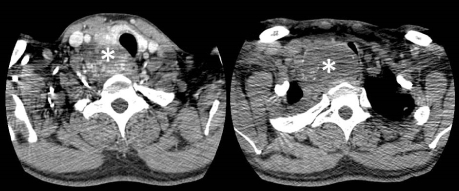
Figure 1. Infiltration of mediastinum with mass effect.
The cervical histopathological study, evidence Follicles of various sizes, in a trabecular pattern, cells present hyperchromatic nuclei and cytoplasm similar to normal follicular cells. (Figure 2a-b), in another field it is possible to observe a papillary pattern. (Figure 2c)
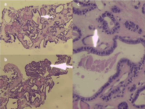
Figure 2. a. Follicles of various sizes, in a trabecular pattern, b. Cells with hyperchromatic nuclei and cytoplasm similar to normal follicular cells, c. Papillary pattern.
It was diagnosed, follicular thyroid carcinoma with metastases to the mediastinal region. The patient dies 4 days after admission to the intensive care unit due to respiratory complication, associated with superior vena cava syndrome.
The follicular thyroid carcinoma has a greater preponderance for the female sex; however, the male sex has also been associated with an advanced age and a large tumor size, with a poor prognosis [4-5] the case presented shows a patient of 43 years of males, which is not consistent with the literature, most of the follicular carcinomas present at premature ages are associated with hereditary syndromes, such as Cowden's disease, Werner's syndrome and the Carney complex [6], we do not count With studies that show if our case belongs to one of the presented syndromes.
It is usually presented as slow enlargement of the thyroid tissue. Histologically it is distinguished from follicular adenoma only by the presence of capsular and vascular invasion [7]. In our anatomopathological study we cannot appreciate the capsular and vascular invasion as we performed a trucut biopsy in the left lateral area to the thyroid gland, obtaining invasive tissue.
Distant metastases are more common in follicular thyroid carcinoma than in papillary thyroid carcinoma (PC) [8]. Distant metastases are common in the lungs and bones [9]. Lymph node metastasis is uncommon. The early stage has a good prognosis (99% survival at 5 years Stage I and II). In stage IV of the disease as presented, survival is 47% to 5 years [10]. In the case presented mediastinal metastasis leading to superior vena cava syndrome is also present, in addition to an emergency with myalgias, arthralgias, cough with hemoptysis associated with respiratory distress, as a cause of tumor compression. Metastasis to lymph nodes is not evident, which is consistent with the literature [10].
The pillar treatment is almost total or total thyroidectomy, with or without neck dissection, followed by suppression of TSH with levothyroxine. A higher stage generally requires radiotherapy with radioactive iodine, occasionally with high doses of up to 200 mCi, to remove remnant local thyroid tissue and distant metastatis[11]. External beam radiation for metastasis is not widely used, since That there are conflicting reports [12]. In the case presented, no tumor-directed treatments were performed because the patient dies because of the superior vena cava syndrome.
One of the rare anatomopathological findings is the combination of more than one histopathological pattern. The combination of more than one different pattern of thyroid cancer is only reported three times. In 2003 by Bohorquez et al. [13] reported a case of a 27-year-old female patient with a combination of a follicular pattern, a medullary pattern And a papillary pattern, clearly separated from each other. Another finding was reported by Kenko-Cupisti [14] in 2005 who found occurrence of three types of thyroid cancer in a recurrent goiter both with a well-located location. And a last report was made by Zerpa de Miliani et al. [15] in 2007 who found three types of thyroid cancer in a patient with recurrent goiter. Our finding differs from the reported cases, in which both the follicular pattern and the papillary pattern were combined. A finding consistent with the study by Bohorquez et al. [13] and ours are the premature ages of both patients; However due to lack of evidence we cannot affirm that this is a phenomenon related to a premature age, more studies are needed to confirm their relationship.
- Mohan H (2012) Patología 6aed. Buenos Aires: Medica Panamericana
- Fletcher K (2013) Diagnostic histopathology of tumors. 2 vols. 4a ed. Philadelphia: Elsevier
- DeLellis R, Lloyd R, Heitz P, Eng C (2004) WHO Classification of Tumours, Pathology and Genetics of Tumours of Endocrine Organs. IARC Press, Lyon: 73-76.
- O'Neill CJ, Vaughan L, Learoyd DL, Sidhu SB, Delbridge LW, et al. (2001) Management of follicular thyroid carcinoma should be individualised based on degree of capsular and vascular invasion. European journal of surgical oncology: the journal of the European Society of Surgical Oncology and the British Association of Surgical Oncology 37: 181-185
- Ito Y, Miyauchi A, Tomoda C, Hirokawa M, Kobayashi K, et al. (2014) Prognostic significance of patient age in minimally and widely invasive follicular thyroid carcinoma: investigation of three age groups. Endocr J 61: 265-271. [Crossref]
- Thompson L (2013) Diagnostic pathology. Head and neck. España: Marban;2013.
- Haigh PI (2002) Follicular thyroid carcinoma. Curr Treat Options Oncol 3: 349-354. [Crossref]
- Grebe SK, Hay ID (1995) Follicular thyroid cancer. Endocrinol Metab Clin North Am 24: 761-801. [Crossref]
- D'Avanzo A, Treseler P, Ituarte PH, Wong M, Streja L, et al. (2004) Follicular thyroid carcinoma: histology and prognosis. Cancer 100: 1123-1129. [Crossref]
- Cooper DS, Doherty GM, Haugen BR, Kloos RT, Lee SL, et al. (2009) Revised American Thyroid Association management guidelines for patients with thyroid nodules and differentiated thyroid cancer: the American Thyroid Association (ATA) guidelines taskforce on thyroid nodules and differentiated thyroid cancer. Thyroid 19: 1167-214.
- Ortak T, Uraloglu M, Uysal AÇ, Orbay H, Tekin F, et al. (2008) Reconstruction of sternal defects with pectoralis major muscle flap. Eur J Plast Surg 30: 223-228
- Mishra A, Mishra SK, Agarwal A, Agarwal G, Agarwal SK (2001) Surgical treatment of sternal metastases from thyroid carcinoma: report of two cases. Surg Today 31: 799-802.
- Bohorquez CL, González-Campora R, Loscertales MC, Escudero AG, Mezquita JC (2003) Solitary fibrous tumor of the thyroid with capsular invasion. Pathol Res Pract 199: 687-690.
- Kenko C, U Ramp R, Donner AW, Krausch M, Eisenberger CF, et al. (2005) Synchronous Ocurrente of follicular, papillary and medullary thyroid carcinoma in recurrent goiter. Endocrine Journal 52: 281-285.
- De Miliani Z, Uzcategui R, Miliani N, Gomez R (2016) Simultaneous occurrence of papillary, follicular and medullary thyroid carcinoma in a patient with recurrent goiter. Rev Venez Endocrinol.