Abstract
Trachyonychia is a nail alteration occurring mainly in children in isolated form or acquired revealing an underlying dermatological pathology.
We report a series of trachyonychia in children diagnosed from 2010 to 2021 in consultation, at the dermatology department of CHU IBN Rochd of Casablanca. Eleven patients with the diagnosis of pediatric trachyonychia were available for follow-up. The mean age of appearance was 6.4 years (range 3–12 yrs). Eleven cases were identified, including 6 girls and 5 boys, the sex ratio was 0.83, the average age was 6.4 years with extremes ranging from 3 to 12 years, the average age of onset was 19 months, the disease was isolated in 8 cases or 72%, associated with psoriasis in 1 patient or 9%, a case of alopecia was noted or 9% and eczema lesions in 1 patient or 9%. Our patients were treated with locally applied dermocorticoids, the evolution was good in 5 patients (45%), stationary in 4 patients (36%) and mild in 2 patients (18%).
Trachyonychia is a basic nail lesion characterized by longitudinal grooves or striations, discoloration, roughness and onychorrhexis and the evolution is variable and can be towards a very progressive improvement spontaneously with age with the possibility of complete cure.
Keywords
trachyonychia, children, clinical aspect, treatment
Introduction
Trachyonychia is a disorder of the nail unit occurring mainly in children that can evolve in isolated form, only affecting the nails or acquired, revealing an underlying dermatological pathology such as peladic dermatitis, lichen planus, or atopic dermatitis, sometimes in the form of isolated involvement. When all the nails are affected, it is called the twenty nails syndrome. Long-term follow-up data on these patients are limited but suggest possible regression of nail findings over time. The application of strong topical corticosteroids or intramatricial injections can accelerate this process. We report a series of trachyonychia cases in children.
Materials and Methods
This is a retrospective study that included all cases of trachyonychia diagnosed from 2010 to 2021 in consultation, at the dermatology department of the CHU IBN Rochd in Casablanca
Results
Eleven cases were identified including 6 girls and 5 boys. The sex ratio was 0.83. The average age was 6.4 years with extremes ranging from 3 to 12 years, the age of onset was on average 19 months. Lesion was isolated in 8 cases (72%) (Figure 1), associated with psoriasis in 1 patient (9%). One case of alopecia areata was observed (9%) as well as eczema lesions in 1 patient (9%). Our patients were treated with topical corticosteroids the outcome was satisfiying in 5 patients (45%) (Figure 2). Stationary in 4 patients (36%) and mild in 2 patients (18%).

Figure 1: Opaque trachyonychia. The nails show a rough surface, longitudinal ridges, and a ‘sandpapered’ appearance
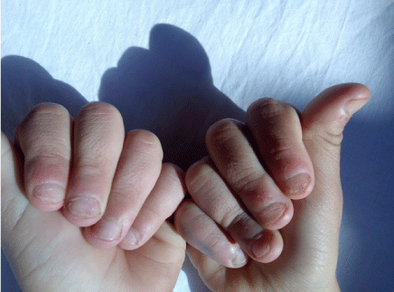
Fig 2 : Good clinical evolution under dermocorticoids
Discussion
Pediatric trachyonychia remains a poorly understood pathology refers as "rough nails", it is characterized by longitudinal striations, ridges, fissures and/or pitting. It can occur at any age, predominant in children with a peak between 3 and 12 years old [1-3]. Trachyonychia can occur idiopathically or in association with other conditions such as alopecia areata, psoriasis, lichen planus, vitiligo and incontinentia pigmenti.
The rough appearance of the nails with excessive longitudinal ridges is characteristic and the cuticles can take on a jagged and thickened appearance [4]. There are two subtypes which have been described on the basis of clinical appearance and severity. The opaque form which remains more severe, characterized by rough nails that appear to have been rubbed with sandpaper and the shiny less severe form, resulting in shiny, opalescent nails with many pitting. This form is frequently associated with alopecia areata.
It is important to think about other differential diagnoses of ungual dystrophy, in particular onychomycosis, hence the interest of a skin-mucous and phaneric examination in search of associated signs, completed by a nail biopsy which is not systematic, but which may be indicated in severe forms or in the event of clinical ambiguity.
The duration of the disease remains shorter in the pediatric population with the possibility of spontaneous recovery, especially in idiopathic forms. It is therefore preferable to ensure supervision and reassure parents. Despite the harmless nature of the disease, patients are requesting treatment because of the esthetic character that can have a negative impact on the quality of life. A therapeutic algorithm can be proposed based on an initial active observation, followed by a trial period of topical corticosteroids, then steroid injections in case of failure of topical treatments, then a systemic treatment with nail biopsy for severe or intractable forms. When the clinical presentation is associated with an underlying disease, treatment of the latter is helpful in improving the appearance of the nails [5].
Conclusion
The interest of our series lies in the importance of recognizing the appearance of trachyonychia and looking for signs indicating an underlying pathology. The diagnosis is often clinical, but a nail biopsy may be suggested to rule out lichen planus or other skin diseases and the treatment is strongly requested in front of the display of the pathology.
References
- Kumar MG, Ciliberto H, Bayliss SJ (2015) Longterm follow-up of pediatric trachyonychia. Pediatr Dermatol 32: 198–200. [Crossref]
- Tosti A, Bardazzi F, Piraccini BM, Fanti PA (1994) Idiopathic trachyonychia (twenty-nail dystrophy): a pathological study of 23 patients. Br J Dermatol 131: 866–872. [Crossref]
- Sehgal VN (2007) Twenty nail dystrophy trachyonychia: an overview. J Dermatol 34: 361–366. [Crossref]
- Chu DH, Rubin AI (2014) Diagnosis and management of nail disorders in children. Pediatr Clin North Am 61: 293–308. [Crossref]
- Haber JS, Manasmon M, Rubin I (2016) Trachyonychia: Review and Update on Clinical Aspects, Histology, and Therapy. Skin Appendage Disorders 2: 109–115. [Crossref]