We describe an asymptomatic 78-year-old man with a unique combination of severe aortic stenosis (AS), left atrial myxoma, biopsy-confirmed transthyretin cardiac amyloidosis and triple vessel coronary artery disease, who had successful resection of the myxoma, aortic valve replacement and triple coronary artery bypass grafting.
cardiac amyloidosis, aortic stenosis, myxoma, cardiac imaging, coronary artery disease, case report
A 78-year-old man, asymptomatic with atrial fibrillation and flutter (Figure 1A) and AS, attended follow up in the valve clinic. Transthoracic echocardiogram showed a small echo- bright mass in the left atrium, along the atrial septum. (Figure 1B).
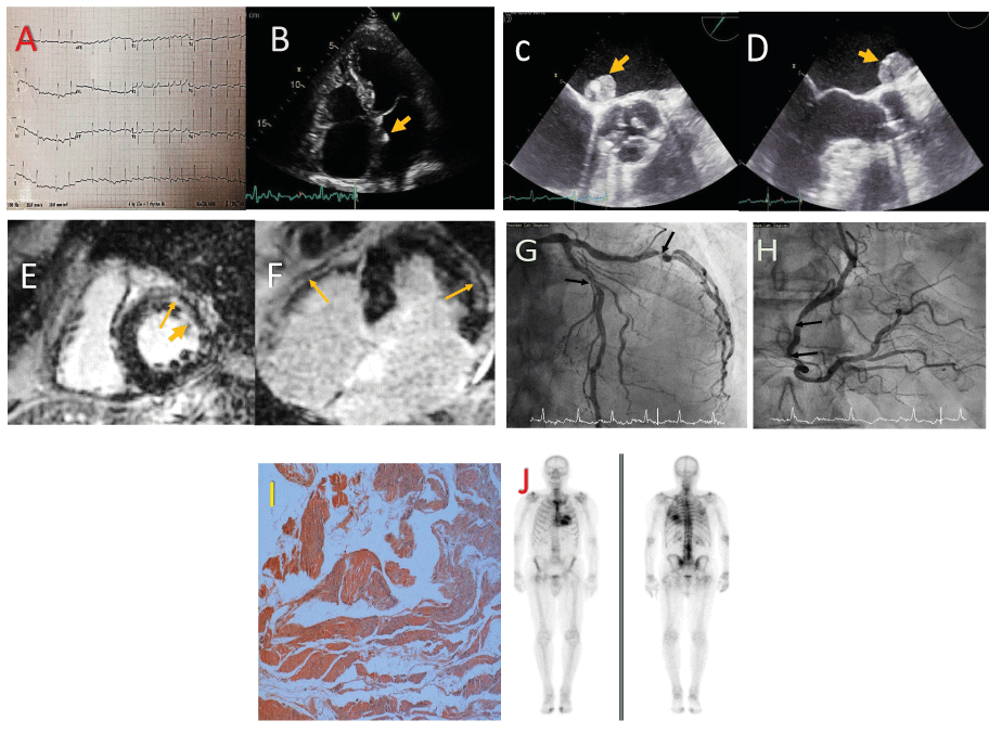
Figure 1.
A. Twelve-lead ECG demonstrating LVH with non-specific ST and T wave abnormalities.
B. Apical4-chamber transthoracicsystolic image demonstrating bi-atrial dilatation and a small echogenic mass attached to the left side of the atrial septum (arrow).
C and D. Transoesophageal short axis image at the base of the heart and long axis image, demonstrating in greater detail the mass attached to the atrial septum (arrow).
E and F - Late gadolinium enhancement on mid-cavity short-axis and on horizontal long axis images demonstrating coarse mid-wall fibrosis of both ventricles (thin arrow) as well as subendocardial fibrosis in the LV (thick arrow).
G and H - Left and right coronary artery angiograms demonstrating diffuse atherosclerosis with areas of focal stenosis in all 3 main branches (arrows) and acceptable distal ‘targets’ for by-pass grafting.
I. Congo red stained section of myocardial biopsy highlighting abnormal protein deposits.x40 magnification.
J. DPD scan showing increased tracer uptake within themyocardium.
Transoesophageal echocardiogram demonstrated severe AS with an aortic valve area on planimetry of 0.8 cm2 with mild central aortic regurgitation, and a 1.4 cm x 1.7 cm mass attached to the left side of the interatrial septum. (Figures C and D) (Clip 1) (Clip 2).
Urgent cardiac magnetic resonance imaging showed LVEF 43%, asymmetrical left ventricular hypertrophy with a maximal wall thickness of 13 mm and severe degenerative AS with AVA 1.0 cm2 on planimetry. There was diffuse circumferential subendocardial and mid- wall LV fibrosis suggestive of amyloidosis and a sessile mass attached to the inferior rim of the fossa ovale in the left atrium, likely a myxoma (Figures E and F).
Preoperative coronary angiogram showed moderate proximal LAD disease, significant proximal LCx disease and moderate diffuse RCA disease. (Figures G and H) (Clip 3)(Clip 4).
The patient underwent an uneventful resection of the left atrial mass together with aortic valve replacement (25mm Perimount bioprosthesis) and coronary artery bypass using 3 grafts. Biopsies confirmed myocardial amyloid deposits (Figure I) and a myxoma. The patient was discharged on postoperative day 10.
Review in outpatients showed excellent recovery from surgery. There was no plasma cell dyscrasia on serum and urine electrophoresis. Technetium-99m (99mTc) and 3,3- diphosphono-1,2-propanodicarboxylic acid (DPD) scan showed early (grade 1-2) cardiac amyloid involvement (Figure J), while serum amyloid P component (SAP) scan showed no visceral amyloid deposits. TTR genetic testing was negative. The patient was doing well clinically and was started on a trial of Transthyretin stabiliser AG 10.
This case illustrates an uncommon combination of cardiac pathologies in a clinically asymptomatic individual in whom regular follow-up for aortic stenosis prompted the diagnosis of left atrial myxoma, coronary artery disease and transthyretin amyloidosis (ATTR). ATTR is often under diagnosed due to the limited specificity of clinical manifestations, and low sensitivity of electrocardiogram and echocardiography [1]. CMR has a high accuracy for diagnosing cardiac amyloidosis with the typical LGE distribution (sensitivity 86 - 88%, specificity 86 - 90%) [2]. Aortic stenosis can coexist with amyloidosis, and the latter should not deter from AVR when clinically indicated [3].
Myxomas are the most common cardiac neoplasm accounting for about 50% of all tumours of the heart [4]. Clinical presentation ranges from no symptoms to constitutional symptoms or to intracardiac obstruction, such as mitral obstruction, and coronary or systemic embolization [5]. Overlying thrombus on the surface of the tumour often embolises rather than the tumour itself [6]. Surgical resection is the only effective treatment to prevent this debilitating and catastrophic complication, [5] with excellent prognosis following excision [4].
- Teng C, Li P, Bae JY, Pan S, Dixon RAF, et al. (2020) Diagnosis and treatment of transthyretin-related amyloidosis cardiomyopathy. Clin Cardiol 43: 1223-1231.[Crossref]
- Vogelsberg H, Mahrholdt H, Deluigi CC, Yilmaz A, Kispert EM, et al. (2008) Cardiovascular magnetic resonance in clinically suspected cardiac amyloidosis: noninvasive imaging compared to endomyocardial biopsy. J Am Coll Cardiol 51: 1022-1030.[Crossref]
- Ternacle J, Krapf L, Mohty D, Magne J, Nguyen A, et al. (2019) Aortic Stenosis and Cardiac Amyloidosis: JACC Review Topic of the Week. J Am Coll Cardiol 74: 2638-2651.[Crossref]
- Nwiloh J, Oludara M, Adebola P (2011) Left atrial myxoma: case report and literature review. East Afr Med J 88: 71-72.[Crossref]
- Amoah AG, Frimpong-Boateng K, Kallen C, Barwasser HM (1998) Atrial myxoma--a case report and review of the literature. West Afr J Med 17: 50-54.[Crossref]
- Latifi AN, Ibe U, Gnanaraj J (2019) A case report of atrial myxoma presenting with systemic embolization and myocardial infarction. Eur Heart J Case Rep 3: ytz104.[Crossref]