With the emergence and spread of Coronavirus Disease 2019 (COVID-19), surgical care of patients has been disrupted for surgeons across the world. Recently several surgical societies are raising concerns about using ultrasonic devices during laparoscopic surgery due to questions regarding a proposed risk of viral transmission of COVID-19. In this review, we will provide an overview of COVID-19's transmission, evaluate available evidence on surgical smoke production and possible risk of viral contamination and discuss the optimization of ultrasonic device use during the ongoing COVID-19 global pandemic.
COVID-19, surgical smoke, ultrasonic, laparoscopic, surgical care, surgery, aerosol formation, virus
A pneumonia of unknown cause detected in Wuhan, China, on December 27th was first reported to the World Health Organization (WHO) Country Office in China on December 31st, 2019 [1]. Early in January, it was determined that these pneumonia cases were due to a novel coronavirus, named SARS-CoV-2, causing a disease called COVID-19.
SARS-CoV-2 is a positive-sense single-stranded RNA virus, whose virion is 60–140 nm in diameter [2]. SARS-CoV-2 has four structural proteins: proteins E (envelope), M (membrane), and S (spike) which create the viral envelope and, through protein S, attach to and fuse with the membrane of a host cell. Protein N (nucleocapsid) holds the RNA genome [2].
SARS-CoV-2 is responsible for a worldwide epidemic, which was declared as a pandemic by the WHO on March 11th, 2020. This pandemic severely limited elective surgical activity throughout the world, as well as triggered multiple surgical societies (e.g., SAGES/EAES, ESGE, AAGL) [3-5] to issue “recommendations” on ways to reduce the risk of virus transmission to the operating room (OR) personnel caring for COVID-19 patients or patients with unknown COVID-19 status. Most of the recommendations are focused on ways to reduce potential exposure via personal protective equipment (PPE), smoke evacuation and potential aerosolization of SARS-CoV-2 by electrosurgical devices or CO2 insufflation of the pneumoperitoneum. These recommendations include setting electrosurgical units to the lowest possible settings for the desired tissue effect and minimizing the use of monopolar electrosurgery, ultrasonic dissectors, and advanced bipolar devices, as they can lead to particle formation in the resulting surgical smoke.
These recommendations were initially focused on laparoscopic surgery, where the risk of viral aerosolization from CO2 insufflation was considered, along with the potential concentration of aerosol created from the use of energy devices.
Concerns have been raised regarding the use of ultrasonic devices where several authors speculated these devices may produce a low-temperature aerosol which do not effectively deactivate the cellular components of virus in patients [6] or may cause more aerosol formation [7]. Even though there is no evidence supporting these theories, different surgical societies, as well as some published literature, are suggesting not to use ultrasonic energy, in favour of electrosurgery.
The aim of this review is to analyse available evidence related to the surgical smoke created by ultrasonic devices as it relates to the risk of COVID-19 transmission during laparoscopic surgery.
The current available evidence suggests the main source of transmission of SARS-CoV-2 is through respiratory droplets (particles > 5-10 μm in diameter) [8] from infected people and through contact with contaminated surfaces [1,9-13].
The detection of viable SARS-CoV-2 in stools of COVID-19 patients, who can shed the virus in the feces for days after respiratory symptoms have disappeared [14,15] and virus RNA has been found in sewage [16,17]. This evidence raises the possibility of fecal-oral transmission, even though there is no data available to support this hypothesis.
Other means of transmission of the virus have not been confirmed. Some studies found the presence of SARS-CoV-2 RNA in blood, but in a very limited number of cases (10-11%) [18,19]. Data on the presence or absence of the infectious virus in blood, plasma or serum has not been reported, as well as blood-borne transmission of COVID-19 disease.
Vertical transmission of the virus from an affected mother to her child seems to be unlikely, even though 4 affected children out of 71 published cases (5.6%) have been observed [20]. While it seems unlikely that this could be a meaningful means of SARS-CoV-2 virus transmission, more data are needed to rule out this mode of transmission.
From a surgical perspective, a large concern is the possibility of conducting a surgical procedure on a patient with unknown COVID-19 status (i.e., false negative, asymptomatic or pre-symptomatic). An increasing number of reports have indicated that some infected persons may not exhibit signs or symptoms of illness, including persons who are presymptomatic (SARS-CoV-2 RNA is detectable before symptom onset) or asymptomatic (SARS-CoV-2 RNA is detectable but symptoms never develop) [21]. The detection of SARS-CoV-2 RNA in presymptomatic or asymptomatic persons does not prove that they can transmit the virus to others. On the other hand, epidemiologic and virologic data seems to suggest the possibility that these subjects might transmit SARS-CoV-2 [22].
As already stated, pulmonary droplets, close contact with affected people and surface contacts are the main accepted means of transmission of SARS-CoV-2 virus.
Airborne transmission is different from droplet transmission as it refers to the presence of microbes within droplet nuclei, which are generally considered to be particles < 5μm in diameter. Airborne particles can remain in the air for longer periods of time than droplets and can be transmitted to others over distances greater than 1 meter. In an analysis of 75,465 COVID-19 cases in China, airborne transmission was not reported [23]. Thus, airborne transmission for SARS-CoV-2 has not yet been clearly established.
Nevertheless, there is some evidence which may indicate aerosol-driven infection. A number of case reports suggest that transmission for asymptomatic individuals in association with normal breathing and talking producing predominantly small droplets [24].
Some studies have demonstrated the presence of SARS-CoV-2 and SARS-CoV-1 (a similar virus from the same family) in aerosol form that lingers in the air and has been reported to travel intra-building and over long distances from the sources in medical and laboratory settings. In a hospital setting, viral RNA has been detected in the air inside COVID-19 patient rooms and in nearby hallways [25]. Another study evaluated SARS-CoV-2 aerosol deposition at 30 sites (i.e., patient areas, medical staff areas, and public areas) in two COVID-19 hospitals in Wuhan, China and found viral RNA in some of the patient areas and in the medical staff protective apparel removal rooms of one of the hospitals, in addition to two public areas (i.e., a department store and a site where the public including outpatients passed by) [26]. All these data seem to support the possibility of droplet nuclei containing virus from infected patients as these studies were conducted in hospitals treating COVID-19 positive patients.
Thus, implications of transmission of viral particles in the environment transported via droplet nuclei is still unknown.
The Royal College of Surgeon of England published an updated General Surgery Guidance on COVID-19 [27] where it warned that laparoscopy should only be considered in selected individual cases where clinical benefit to the patient substantially exceeds the risk of potential viral transmission to surgical and OR teams.
This recommendation was issued in a moment where no data existed on the presence or absence of SARS-CoV-2 virus in peritoneal tissues or organs and was based on the assumption the pneumoperitoneum would have a concentrated amount of aerosol which could contain virus particles and resulting leaks of CO2 may transmit virus to the OR team.
On the other hand, experience with previous coronaviruses was available at the time the RCS Guidance was published. To, et al. [28] published a report investigating the tissue and cellular tropism of SARS-CoV, a predecessor of SARS-CoV-2, in fatal SARS cases by in situ hybridization in six autopsies. These authors found that lung sections from three cases and small intestinal sections from four cases were all positive for viral culture, while all samples were positive for in-situ hybridization (ISH). Other tissue samples, including the heart, lymph nodes, bone marrow, muscles, and organs contained in the abdominal cavity, such as liver, spleen, kidney, were all culture- and ISH-negative. These results confirmed that the lung and the intestine were the only two organs where presence of SARS-CoV was confirmed.
Two case reports investigating the presence of SARS-CoV-2 in the peritoneal fluid from COVID-19 patients were recently published. Ngaserin et al. [29] published a case of a 21-year old male with no pre-existing co-morbidities under active quarantine as part of a known cluster of COVID-19 with acute pain in the right iliac fossa and vomiting. A nasopharyngeal COVID-19 polymerase chain reaction (PCR) swab test was positive, and the patient underwent laparoscopic appendectomy. Upon entry in the abdominal cavity, 5 ml of seropurulent peritoneal fluid from the Morrison’s pouch, right paracolic gutter and pelvis was aspirated and sent for COVID-19 PCR. After appendectomy, peritoneal washings were also collected prior to the end of the case and sent for COVID-19 PCR. The peritoneal fluid samples on entry and just before extraction of the appendix were both found to be negative for COVID-19 by PCR.
Coccolini et al. [30] published a case of a 78-year-old man who presented for abdominal pain associated with alteration of the bowel function. He had fever, cough and mild respiratory symptoms; the respiratory nasal swab was positive for SARS-CoV-2. The patient was admitted with a diagnosis of intestinal mechanical obstruction due to small bowel volvulus associated to SARS-CoV-2 pneumonia. While conducting a laparotomy, it was found that the volvulus was due to an omental band attached to the right iliac fossa. The band was dissected, and no bowel resection was performed. Two swabs were obtained from peritoneal fluid and sent for SARS-CoV-2 detection. The authors demonstrated the presence of a high concentration of viral RNA, but virus isolation - which would have provided stronger evidence of infectivity - could not be performed. Notwithstanding the demonstration of the presence of the sole RNA, the authors strongly commented the surgical procedure should have been considered as a risk of infection to the OR team.
The available evidence are inconclusive and contrasting. The presence of a transmittable form of SARS-CoV-2 in the peritoneal cavity has not been demonstrated and thus the presence of virus in the aerosol of the pneumoperitoneum still remains a hypothesis.
Energy devices, including electrosurgery devices, ultrasonic devices, lasers and high-speed drills, burrs and saws, all produce surgical smoke. Surgical smoke is a gaseous by-product produced during surgical procedures and is referred also as aerosol, cautery smoke, diathermy plume, plume, or smoke plume.
Surgical smoke contains approximately 95% water or steam, and 5% particulate matter from cellular debris and chemical compounds in the gaseous phase [31,32]. Data on the morphology, size, and composition of surgical smoke are scant [33-35].
There are significant differences in how ultrasonic and electrosurgical instruments cut and coagulate tissues and, thus, in how they produce surgical smoke. The ultrasonic device denatures protein by the transfer of mechanical energy to the tissues that is sufficient to break hydrogen bonds and by the generation of heat from friction that results from the interaction of the blade with the tissue. During this time, the blade becomes warm to the touch [36]. In his initial experiment ultrasonic blades (i.e., no clamp arm), Amaral [36] observed that heat generated in the tissues as a result of stress and friction is limited. In that study, a personal communication to the author indicated that thermographic analysis has demonstrated that ultrasonically activated coagulation does not heat tissues above 80°C. As a result, tissues do not desiccate from the loss of moisture, and they do not burn.
Different recommendations and articles on the use of energy devices in laparoscopy during the COVID-19 pandemic argue against the use of ultrasonic scalpel, based on the belief that this mode of energy is causing higher amounts of surgical smoke, which is also believed to be cooler and, thus, possibly not able to inactivate the virus [6,37].
It is quite difficult to compare the amount of actual smoke volume production from different energy sources, but there are several studies evaluating the amount of smoke particles produced when using electrosurgical or ultrasonic devices.
Ott et al. [38] determined the distribution and concentration of aerosol particles caused by an ultrasonic scalpel with different end effectors (ball, hook and blade) during simulated surgical use with the monitor probe mounted at fixed distances ranging from 10 to 30 cm from the site of aerosol production. In addition, they compared ultrasonic to a monopolar device, and also evaluated the effect of a smoke evacuation system on particle concentration. The interaction of ultrasonically activated devices with tissue produces a biphasic bioaerosol composed of tissue particles and a blood aerosol [38] and a minimal smoke production has been reported for this device [39]. Ott et al. [38] found that fatty tissue generated more particles than lean tissue, but more interestingly, that the local exhaust ventilation smoke-evacuation system dramatically reduced particle concentration exposure and that monopolar electrosurgery (30 W) showed a four-fold increase in particle concentration.
Weld et al. [40] characterized the smoke produced by four energy-based laparoscopic instruments, bipolar macroforceps, ultrasonic scalpel, floating ball, and monopolar shears applied in vitro to porcine psoas muscle. Bipolar energy produced the smallest number of large particles, while bipolar energy and the ultrasonic scalpel both created a relatively small number of small particles (Table 1). In contrast, the standard monopolar scissors and the floating ball device both created a large number of both small (< 500 nm) and large (> 500 nm) particles (Table 1). In the same study, the authors demonstrated that bipolar and ultrasonic devices had the least effect on visibility by the smoke particles generated.
Table 1. Concentration of particles in surgical smoke from several different energy devices (Modified from Weld)
|
Concentrations of particles (per cm3) |
|
Particles < 500 nm |
Particles > 500 nm |
Total particles |
Bipolar |
5.3 × 10⁵ |
869 |
5.36 × 10⁵ |
Ultrasonic |
6.10 ×10⁵ |
1.48 × 10³ |
6.11 × 10⁵ |
Floating Ball |
1.65 × 10⁷ |
6.61 × 10³ |
1.65 × 10⁷ |
Monopolar |
4.40 ×10⁷ |
8.13 × 10³ |
4.40 × 10⁷ |
Background control |
3.86 ×10³ |
17 |
3.88 × 10³ |
* Data regarding particles generated by advanced bipolar devices was not available in the scientific literature at the time of writing.
Another study from Lamberton et al. [41] measured particulates in vapor of ultrasonic scalpel and three other advanced bipolar devices using a laser photometer at a distance of 5 cm. They demonstrated that the Ultrasonic Scalpel produced the least amount of smoke (mean 2.88 ppm versus 74.1 ppm and 21.6 ppm, p = 0.0001) of two other devices and similar (mean 12.5 ppm, p = 0.11) of the third advanced bipolar.
From this evidence, it can be concluded that smaller particles (mainly produced by monopolar devices) which have higher concentrations remained in suspension longer due to their lighter mass, which increased the visual obstruction of plume, while larger particles (mainly produced by ultrasonic devices) dissipated more quickly because of their greater mass. More obstruction could suggest a higher concentration of plume. Moreover, it does not seem that ultrasonic produces more smoke but, on the contrary, it may be even less.
Another opinion that has been suggested is that ultrasonic produces a cooler surgical smoke than electrosurgical devices. Although a number of articles report this information [37,42-47] none of them performed or referenced a scientific experiment demonstrating this statement. The articles all point back to the original work from Amaral, cited above, where the author reports a personal communication about the fact that thermographic analysis would have demonstrated that ultrasonically activated coagulation does not heat tissues above 80°C [36].
A recent study by Hayami et al. evaluated the temperature of the steam from an ultrasonic shear and compared it to an advanced bipolar device [48]. These authors performed an ex vivo animal study using porcine muscle and tested the devices in four different combinations of device and muscle conditions, including dry–dry, dry–wet, wet–dry, and wet–wet. In addition, grasping range (proportion of the length of tissue held between the jaw of devices to that of the jaw) changed to 1/3, 2/3, and 3/3, under each condition.
The temperatures of energy devices and steam were measured using thermography at 0-, 1-, 3-, 5-, and 10-mm away from the energy device. Although the maximum temperature of the devices was significantly higher with ultrasonic shears than with advanced bipolar, the maximum temperature of steam was significantly higher with the latter, in almost all situations. It should be stressed though, that ultrasonic devices reached the critical temperature of 60°C (the study was performed to evaluate possible thermal damage to nervous structures) 1 mm away from the device using a 1/3 grasp for the wet/wet and wet/dry condition and that also advanced bipolar did not reach this critical temperature in many situations at the same distance. Moreover, the advance bipolar, as well as ultrasonic shears, did not reach 600°C at distances > 1 mm.
The authors acknowledged a number of limitations of the study [48]. The experimental nature of the study could not reflect its application to humans and the porcine tissues may not have accurately reflected the in vivo setting, mainly due to the lack of a blood supply. Furthermore, the starting temperature of the experiment (significantly cooler than clinical settings) might have had a significant impact on the temperature of energy device. The temperature and the direction of steam from the energy device may have been dependent on the grasping range, the volume of the moisture content between the jaws, and the distribution of the heat source at the contact surface of the jaws. Lastly, a pattern of which direction the steam goes in and the influence of several conditions, including pneumoperitoneum or gravity/patient position, on it are very important.
A study which showed opposite results was performed by Emam et al. [49], who compared two ultrasonic devices at the three power settings (3, 4, and 5) in random fashion in an experimental setting in pigs. Thermography was used for real-time mapping of the heat production during use of the ultrasonic shears and it was observed that the zone around the jaws that exceeded 60°C with continuous ultrasonic dissection for 10 to 15 seconds at level 5 measured 25.3 and 25.7 mm for the two different ultrasonic devices. At this power setting and an activation time of 15 seconds, the temperature 1.0 cm away from the tips of the instrument exceeded 140°C. All results are reported in Table 2. It can be observed that temperatures 1 cm away from the jaws of both instruments were all above 80°C for power settings level 4 and level 5.
Table 2. Peak temperatures after 5, 10, and 15 seconds activation. (Modified from Emam)
|
Peak temperature - °C (SD) |
|
Activation |
Ultrasonic |
Autosonix |
P value |
|
Jaws |
1 cm away |
Jaws |
1 cm away |
Jaws |
1 cm away |
5 secs |
|
|
|
|
|
|
Level 3 |
69.0 (14.7) |
48.7 (10.3) |
73.9 (13.3) |
51.7 (9.3) |
NS |
NS |
Level 4 |
118 (31.3) |
83.1 (21.9) |
123 (34.1) |
86.7 (23.7) |
NS |
NS |
Level 5 |
124.2 (57.9) |
91.4 (7.8) |
127 (57.6) |
98.7 (9.4) |
NS |
NS |
10 secs |
|
|
|
|
|
|
Level 3 |
74.4 (20.4) |
52.2 (12.4) |
82.9 (17.4) |
55.6 (12.2) |
NS |
NS |
Level 4 |
120.1 (36.0) |
89.4 (23.6) |
125.3 (35.0) |
94.2 (23.0) |
NS |
NS |
Level 5 |
192.3 (44.9) |
134.6 (31.5) |
197.6 (50.2) |
138.3 (35.3) |
NS |
NS |
15 secs |
|
|
|
|
|
|
Level 3 |
74.8 (12.3) |
52.3 (8.6) |
86.3 (11.6) |
60.4 (8.1) |
NS |
NS |
Level 4 |
136.4 (36.1) |
95.5 (25.3) |
148.4 (33.1) |
103.9 (23.2) |
NS |
NS |
Level 5 |
206.9 (27.9) |
144.8 (27.9) |
211.3 (42.7) |
147.9 (29.9 |
NS |
NS |
The limited and contrasting evidence around the temperature of surgical smoke produced by ultrasonic devices vary and are difficult to interpret, necessitating the need for more clinical studies to address this question.
The potential dangers related to the inhalation of surgical smokes has been a question pondered dating long before the COVID-19 pandemic. Indeed, Eubanks et al. [50] published a study evaluating measures to reduce the risk of human immunodeficiency virus (HIV) transmission during laparoscopic procedures almost 30 years ago.
There have been reports of varying infectious material detected in surgical smoke. A recent systematic review investigating the potential risks to the OR team discussed six separate studies which assessed the presence of infectious material in surgical smoke [47]. All six of the studies reviewed were on CO2 laser-produced smoke, except for one that compared laser with electrocautery [51]. One study confirmed the potential for bacterial cell culture growth in laser-derived smoke [52], demonstrating that 5 of 13 participants showed a growth of coagulase-negative Staphylococcus, one of the five samples grew Corynebacterium, and one grew Neisseria. Another study identified human papillomavirus (HPV) DNA in the surgical smoke of plantar warts [51]. During this study, five of eight laser-derived vapors and four of seven electrocoagulation-derived vapors tested positive for HPV DNA, but greater amounts of HPV DNA were recovered in the laser vapor than in the electrocoagulation vapor [51]. Other studies failed to demonstrate the presence of HPV in the laser smoke or the contagion of cells cultured with laser smoke [52-56].
Only one study has evaluated viral emission in a clinical laparoscopic setting [57]. Hepatitis B virus (HBV) was isolated in surgical smoke from electrosurgical devices in HBV-positive patients undergoing laparoscopy. In 10 of 11 cases, there was detectable HBV in the collected smoke. Two studies evaluated the presence of HIV-related molecules in smoke from energy devices. One in vitro study [58] assessed the use of laser to vaporize HIV-positive cultured cells and demonstrated that cultures of the silastic collection tubing revealed p24 HIV gag antigen (3/12 tube segments at the end of 1 week and 1/12 tube segments at 2 weeks). No sustained infection of HIV cultured cells was observed at the 28th day. PCR analysis of particulate debris obtained from the silastic collection tubing was positive from proviral HIV DNA in samples which were immediately taken and on day 14. The other in vitro study [59] found cell infection detected by the appearance of HIV-1 P-24 core antigen in cell cultured with cool aerosols and vapors generated by a 30,000 RPM spinning router tip, an instrument similar to an oscillating bone saw. It should be highlighted that this type of device is completely different from ultrasonic scalpels, which have a frequency of 55,500 Hz as opposed to 500 Hz (= 30,000 RPM) of the oscillating bone saw used in the study.
The only evidence supporting transmission of disease via surgical smoke was in an open procedure using CO2 to treat genital warts, conferring HPV to several members of the OR team [60-61].
As can be noted, there are data on the presence of infectious material in the smoke from ultrasonic devices in either an open setting or in minimally invasive approach.
There are contrasting data on the presence of viable cells in the smoke from ultrasonic shears. One study demonstrated, in an experimental setting, that large quantities of cellular debris were trapped in the plume from both ultrasonic hook and monopolar with a needle probe (cutting mode at 60 W) after ablation of tumors. However, no viable cells were isolated from the smoke of either device [62]. On the other hand, In, et al. [63] compared the presence of cancerous cells in the smoke of different energy devices (electrocautery, radiofrequency ablation and ultrasonic scalpels) and found viable cells in smoke retrieved from a distance of 5 cm only from the ultrasonic scalpel; at 10 cm distance the rate was much lower. There were no viable cells in surgical smoke from the electro-surgical unit or radiofrequency ablation device [63]. Tumor growth was seen in total 16 of 40 injection sites in animals. All tumors contained highly mitotic cells including irregularly shaped nuclei, consistent with malignant tumours [63].
All recommendations issued to address the risk of OR staff virus transmission put an emphasis on the use of appropriate personal protective equipment, as well as the adoption of smoke evacuation systems [3-5].
In particular, when aerosol generating procedures are conducted on patients suspected or positive for COVID-19 and when assisting on procedures on airways in all patients, surgeons are advised to wear PAPR (Powered Air Purifying Respirator) or N95 respirator plus face shield or eye protection, gown, and double gloves [64]. When caring for patients suspected or positive for COVID-19, eye protection/face shield, gown and gloves should also be worn, but a surgical mask may also be suitable [64].
An N95 mask or respirator is a particulate-filtering tool that meets the U.S. National Institute for Occupational Safety and Health (NIOSH) standards and are classified to filter at least 95% of airborne particles >300nm [65]. N95 respirators are considered functionally equivalent to certain respirators regulated under non-U.S. jurisdictions, such as FFP2 respirators of the European Union and KN95 respirators of China. In contrast, surgical masks are intended to provide a barrier to splashes and droplets that may impact the wearer’s nose, mouth and respiratory tract. They do not provide protection against airborne (aerosol) particles [66].
Smoke evacuation systems are designed to reduce surgical smoke for improved visibility and a cleaner OR environment. They are normally equipped with High-Efficiency Particulate Air (HEPA) or, more frequently, with greater performing Ultra-Low Particulate Air (ULPA) filter. HEPA filters have a minimum 99.97% efficiency rating for removing particles greater than or equal to 300 nm [67], while ULPA filters can remove from a minimum of 99.999% of airborne particles with a maximum particle penetration size of 50-120 nm [68].
Currently, it is assumed that SARS-CoV-2 is transmitted as it attaches to larger respiratory water droplets that are in excess of 20,000 nm [69]. Additionally, when SARS-CoV-2 is aerosolized in a CO2 gas suspension the droplet size is assumed to be less than 10,000 nm in size [69]. As mentioned previously, it is unknown if SARS-CoV-2 is contained in surgical smoke and thus the actual particle size, if it exists, is unknown. But, if intact viable virus is attached to a water droplet found in surgical smoke it is reasonable to assume that it will be captured and filtered by an N95 mask, as well as both HEPA and ULPA filters, as shown in figure 1.
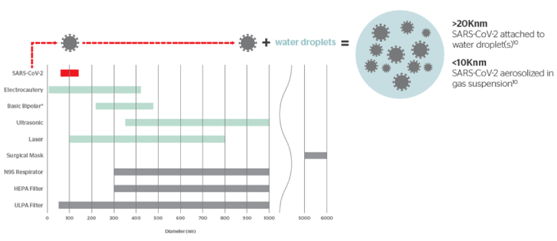
Figure 1. Relative diameters of surgical smoke particles compared to SARS-CoV-2 and commonly used masks and filters
Given the potential, yet not demonstrated, risk with SARS-CoV-2 being present in surgical smoke, a smoke evacuation system should be used. The diligent use of a smoke evacuation system with a high-efficiency filter has been identified as a feasible and potentially useful way for surgical smoke to be reduced [38,70]. The smoke evacuator should be activated at all times when airborne particles are produced during all surgical or other procedures.
Other measures can be taken to reduce the risk of escape of aerosol from the pneumoperitoneum. In particular, incisions for ports should be as small as possible to allow for the insertion of ports but not allow for leakage around ports, the pressure in the pneumoperitoneum should be as low as possible for the desired effect to minimize CO2 leakage from the trocar, reduce the number of times instruments are removed from the trocar to minimize leakage of CO2 from the pneumoperitoneum (e.g., by taking advantage of the multifunctional nature of an advanced energy device, and in particular of ultrasonic) can reduce leakage of CO2 due to reduced instrument exchanges and all pneumoperitoneum should be safely evacuated via a filtration system before closure, trocar removal, specimen extraction or conversion to open [3]. There is no evidence to date that balloon trocars are more effective at reducing CO2 leakage, as they are primarily designed to improve retention in the abdomen [70].
The COVID-19 pandemic has put an unprecedented amount of pressure on healthcare systems worldwide, causing a temporary pause of elective surgical procedures, as well as the need to adopt strategies to reduce the risks of viral transmission for both patients and healthcare professionals.
In this challenging scenario, surgical societies felt the need to take immediate action to define ways to protect OR teams who are caring for suspected or confirmed COVID-19 patients. For these reasons, recommendations were issued which are based mainly upon potential risks, but not exactly on the available evidence. This resulted in recommendations to completely avoid laparoscopy in favor of laparotomy in the face of potentially higher exposure risks due to the concentration of aerosol in the pneumoperitoneum. Fortunately, this recommendation was short-lived after the surgical societies reflected on the scant evidence regarding the risks of transmission of SARS-CoV-2. Instead, they shifted their focus on the real risks of performing all surgeries with an open approach in the face of completely abandoning the significant patient advantages of minimally invasive surgery.
Some of these recommendations, as well as a number of published literature, seem to argue against the use of ultrasonic shears, claiming this type of energy device produces cooler smoke where more viable viral particles could be present, thus increasing the risk of virus transmission. Given the evidence discussed in this review, it is clear that there is no objective evidence to neither confirm these opinions, nor suggest other energy devices as a lower risk or safer.
In summary, the available evidence suggests that:
• The main ways of transmission of SARS-CoV-2 are through respiratory droplets and through contact with contaminated surfaces.
• Other means of transmissions, such as through aerosol, oro-fecal, and vertical (materno-fetal) have been hypothesized but not confirmed.
• Even though SARS-CoV-2 has been detected in aerosolized form in the environment, the implications for transmission are unknown.
• The presence of an intact, transmissible and infective form of SARS-CoV-2 in the peritoneal cavity has not been found. Indeed, only viral RNA was found in one case report and no viral material in the other two.
• It has not been demonstrated that ultrasonic devices produce more smoke, but rather that, in a study, they produced less smoke than monopolar. Ultrasonic devices do produce larger particles that dissipate more quickly, demonstrating greater visibility than monopolar. Larger particle sizes are also more easily filtrated by N95 masks and smoke evacuation systems.
• The limited evidence suggests the aerosol from ultrasonic devices may be cooler than the smoke from bipolar. But this difference was only shown in some conditions tested and only within 1 mm from the tip of the instrument. Beyond that 1 mm distance both ultrasonic and bipolar devices produced smoke and/or aerosol cooler than 60°C, which is considered the lowest temperature responsible for producing irreversible cellular damage.
• There is evidence indicating that some viruses (HIV, HBV, HPV) can be present in the smoke of energy devices. Nevertheless, most of the studies were on open procedures using a CO2 laser, only a few focused on electrosurgical devices and none on ultrasonic shears.
• The only evidence to date illustrating viral transmission through surgical smoke is via the CO2 laser used in an open approach while treating vaginal warts. There is no data to date demonstrating the transmission of any infectious disease through surgical smoke to the OR team during minimally invasive approaches.
It is understandable that a situation like the COVID-19 pandemic would initiate heightened caution and precaution. Our community of healthcare professionals tend to be more conservative for the sake of patient safety, as well as the healthcare professionals who care for them. But we also have a responsibility to make recommendations and decisions based on the entirety of the available data. Care should be taken to not convey inaccurate, and potentially dangerous, opinions not based on evidence. Rather, we feel strongly that it is task of the medical community to provide balanced evidence-based recommendations which highlight the “knowns” and the “unknowns” of the topic under evaluation.
In this regard, we echo the SAGES and EAES recommendations [3] over the use of energy devices. When using monopolar electrosurgery, laser, ultrasonic dissectors and advanced bipolar devices, minimize the creation of surgical smoke. If available, energized devices with attached smoke evacuators should be used.
— Minimize activating any energy devices in a fluid environment.
— Minimize the length of time for a given activation of an energy device and allow the device to cool down between activations.
— For devices with jaws, avoid forcing too much tissue into the jaws.
— Energy devices should be set to the lowest possible power setting for the desired tissue effect.
We should also leverage the increased awareness around the need for a safer laparoscopic environment to introduce the routine use of smoke evacuation and CO2 filtering in all laparoscopic procedures, as well as to set up specific research strategies to increase the evidence that are still missing.
Giovanni A. Tommaselli, Crystal Ricketts, Jeffrey Clymer, and Raymond Fryrear, II are employees of Johnson & Johnson Medical Devices.
- Li Q, Guan X, Wu P, Wang X, Zhou L, et al. (2020) Early Transmission Dynamics in Wuhan, China, of Novel Coronavirus–Infected Pneumonia. N Engl J Med 382 :1199-1207. [CrossRef]
- Zhu N, Zhang D, Wang W, Li X, Yang B, et al. (2020) A Novel Coronavirus from Patients with Pneumonia in China, 2019. N Engl J Med 382: 727-733. [CrossRef]
- Francis N, Dort J, Cho E, Feldman L, Keller D, et al. (2020) SAGES and EAES Recommendations for Minimally Invasive Surgery During COVID-19 Pandemic. Surg Endosc 34: 2327-2331. [CrossRef]
- Americal Association of Gynecologic Laparoscopist (2020) Joint Statement on Minimally Invasive Gynecologic Surgery During the COVID-19 Pandemic. [Accessed: May 7, 2020] Available at: https://www.aagl.org/news/covid-19-joint-statement-on-minimally-invasive-gynecologic-surgery/.
- European Society of Gynecological Endoscopists (2002) ESGE Recommendations on Gynaecological Laparoscopic Surgery during Covid-19 Outbreak. [Accessed May 13, 2020] Available at: https://esge.org/wp-content/uploads/2020/03/Covid19StatementESGE.pdf.
- Zheg MH, Boni L, Fingerhut A (2020) Minimally Invasive Surgery and the Novel Coronavirus Outbreak: Lessons Learned in China and Italy. Ann Surg 272: e5-e6. [CrossRef]
- Balibrea JM, Badia JM, Perez IR, Antona EM, Peña EA, et al. (2020) [Surgical Management of Patients With COVID-19 Infection. Recommendations of the Spanish Association of Surgeons]. Cir Esp 98: 251-259. [CrossRef]
- World Health Organization. (2014) Infection prevention and control of epidemic- and pandemic-prone acute respiratory infections in health care. Geneva: World Health Organization; 2014 [Accessed May 13th, 2020] Available from: https://apps.who.int/iris/bitstream/handle/10665/112656/9789241507134_eng.pdf?sequence=1.
- Liu J, Liao X, Qian S, Yuan J, Wang F, et al. (2020) Community transmission of severe acute respiratory syndrome coronavirus 2, Shenzhen, China, 2020. Emerg Infect Dis 26:1320-1323. [CrossRef]
- Chan J, Yuan S, Kok K, To K, Chu H, et al. (2020) A familial cluster of pneumonia associated with the 2019 novel coronavirus indicating person-to-person transmission: a study of a family cluster. Lancet 395: 514-523. [CrossRef]
- Huang C, Wang Y, Li X, Ren L, Zhao J, et al. (2020) Clinical features of patients infected with 2019 novel coronavirus in Wuhan, China. Lancet 395: 497–506. [CrossRef]
- Burke RM, Midgley CM, Dratch A, Fenstersheib M, Haupt T, et al. (2020) Active monitoring of persons exposed to patients with confirmed COVID-19 — United States, January–February 2020. MMWR Morb Mortal Wkly Rep 69: 245-246. [CrossRef]
- World Health Organization. 2020 Report of the WHO-China Joint Mission on Coronavirus Disease 2019 (COVID-19) 16-24 February 2020 [Internet]. Geneva: World Health Organization; 2020 [Accessed May 13, 2020] Available from: https://www.who.int/docs/default-source/coronaviruse/who-china-joint-mission-on-covid-19-final-report.pdf.
- Wang W, Xu Y, Gao R, Lu R, Han K, et al. (2020) Detection of SARS-CoV-2 in Different Types of Clinical Specimens. JAMA 323: 1843-4. [CrossRef]
- Wu Y, Guo C, Tang L, Hong Z, Zhou J, et al. (2020) Prolonged presence of SARS-CoV-2 viral RNA in faecal samples. Lancet Gastroenterol Hepatol 5: 434–435. [CrossRef]
- Medema G, Heijnen L, Elsinga G, Italiaander R, Brouwer A (2020) Presence of SARS-Coronavirus-2 in Sewage. Medrxiv. doi: https://doi.org/10.1101/2020.03.29.20045880
- Ahmed W, Angel N, Edson J, Bibby K, Bivins A, et al. (2020) First confirmed detection of SARS-CoV-2 in untreated wastewater in Australia: a proof of concept for the wastewater surveillance of COVID-19 in the community. Sci Total Environ 728: 138764. [CrossRef]
- Chen W, Lan Y, Yuan X, Deng X, Li Y, et al. (2020) Detectable 2019-nCoV Viral RNA in Blood Is a Strong Indicator for the Further Clinical Severity. Emerg Microbes Infect 9: 469-473. [CrossRef]
- Peng L, Liu J, Xu W, Luo Q, Chen D, et al. (2020) SARS-CoV-2 Can Be Detected in Urine, Blood, Anal Swabs, and Oropharyngeal Swabs Specimens. J Med Virol. Online ahead of print. [CrossRef]
- Lamouroux A, Attie-Bitach T, Martinovic J, Leruez-Ville M, Ville Y (2020) Evidence for and against vertical transmission for SARS-CoV-2 (COVID-19). Am J Obstet Gynecol 223: 91. [CrossRef]
- Wang Y, Liu Y, Liu L, Wang X, Luo N, et al. (2020) Clinical outcome of 55 asymptomatic cases at the time of hospital admission infected with SARS-Coronavirus-2 in Shenzhen, China. J Infect Dis 221: 1770-1774. [CrossRef]
- Furukawa NW, Brooks JT, Sobel J (2020) Evidence Supporting Transmission of Severe Acute Respiratory Syndrome Coronavirus 2 While Presymptomatic or Asymptomatic. Emerg Infect Dis 26. [CrossRef]
- Ong SW, Tan YK, Chia PY, Lee TH, Ng OT, et al. (2020) Air, surface environmental, and personal protective equipment contamination by severe acute respiratory syndrome coronavirus 2 (SARS-CoV-2) from a symptomatic patient. JAMA 323: 1610-1612. [CrossRef]
- Anderson EL, Turnham P, Griffin JR, Clarke CC (2020) Consideration of the Aerosol Transmission for COVID-19 and Public Health. Risk Anal 40: 902-907. [CrossRef]
- Santarpia JL, Rivera DN, Herrera V, Morwitzer MJ, Creager H, et al. (2020) Transmission potential of SARS-CoV-2 in viral shedding observed at the university of Nebraska medical center. medRxiv. https://doi.org/10.1101/2020.03.23.20039446.
- Liu Y, Nin Z, Chen Y, Guo M, Liu Y, et al. (2020) Aerodynamic analysis of SARS-CoV-2 in two Wuhan Hospitals. Nature. Online ahead of print. [CrossRef]
- Royal College of Surgeons of England. 2020 Updated General Surgery Guidance on COVID-19. 2020. [Accessed May 13, 2020]. Available at: https://www.rcseng.ac.uk/-/media/files/rcs/coronavirus/2nd-update-intercollegiate-general-surgery-guidance-on-covid19-6-april.pdf.
- To KF, Tong JH, Chan PK, Au FW, Chim SC, et al. (2004) Tissue and cellular tropism of the coronavirus associated with severe acute respiratory syndrome: an in‐situ hybridization study of fatal cases. J Pathol 202 :157-63. [CrossRef]
- Ngaserin SH-N, Koh FH, Ong B-C, Chew M-H (2020) COVID-19 Not Detected in Peritoneal Fluid: A Case of Laparoscopic Appendicectomy for Acute Appendicitis in a COVID-19-infected Patient. Langenbecks Arch Surg 405: 353-355. [CrossRef]
- Coccolini F, Tartaglia D, Puglisi A, Lodato M, Chiarugi M (2020) SARS-CoV-2 is present in peritoneal fluid in COVID-19 patients. Ann Surg. Online ahead of print.
- Ulmer BC (2008) The hazards of surgical smoke. AORN J 87: 721-734. [CrossRef]
- Fencl JL (2017) Guideline implementation: surgical smoke safety. AORN J 105: 488-497. [CrossRef]
- Barrett WL, Garber SM (2003) Surgical smoke: A review of the literature. Is This Just a Lot of Hot Air? Surg Endosc 17: 979. [CrossRef]
- Heinsohn P, Jewett DL, Balzer L, Bennett CH, Seipel P, et al. (1991) Aerosols created by some surgical power tools: Particle size distribution and qualitative hemoglobin content. App Occup Environ Hyg 6: 773.
- DesCoteaux JG, Picard P, Poulin EC, Baril M (1996) Preliminary study of electrocautery smoke particles produced in vitro and during laparoscopic procedures. Surg Endosc 10: 152. [CrossRef]
- Amaral JF (1994) The Experimental Development of an Ultrasonically Activated Scalpel for Laparoscopic Use. Surg Lap Endoscop 4: 92-99. [CrossRef]
- Kimming R, Verheijen RHM, Rudnicki M (2020) Robot assisted surgery during the COVID-19 pandemic, especially for gynecological cancer: a statement of the Society of European Robotic Gynaecological Surgery (SERGS). J Gynecol Oncol 31: e59. [CrossRef]
- Ott DE, Moss E, Martinez (1998) Aerosol Exposure from an Ultrasonically Activated (Harmonic) Device. J Am Assoc Gynecol Laparosc 5: 29-32. [CrossRef]
- Tulandi T, Chan KL, Arseneau J (1994) Histopathological and adhesion formation after incision using ultrasonic vibrating scalpel and regular scalpel in the rat. Fertil Steril 61: 548-550. [CrossRef]
- Weld KJ, Dryer S, Ames CD, Cho K, Hogan C, et al. (2007) Analysis of Surgical Smoke Produced by Various Energy-Based Instruments and Effect on Laparoscopic Visibility. J Endourol 21: 347-351. [CrossRef]
- Lamberton GR, His RS, Jin DH, Lindler TU, Jellison FC, et al. (2008) Prospective Comparison of Four Laparoscopic Vessel Ligation Devices. J Endourol 22: 2307-12. [CrossRef]
- Limchantra IV, Fong Y, Melstrom KA (2019) Surgical Smoke Exposure in Operating Room Personnel: A Review. JAMA Surg 154: 960-967. [CrossRef]
- Fan JK, Chan FS, Chu KM. (2009) Surgical smoke. Asian J Surg 32: 253-7. [CrossRef]
- Barrett WL, Garber SM (2003) Surgical Smoke: A Review of the Literature. Is This Just a Lot of Hot Air? Surg Endosc 17: 979-87. [CrossRef]
- Fitzgerald JE, Malik M, Ahmed I (2012) A single-blind controlled study of electrocautery and ultrasonic scalpel smoke plumes in laparoscopic surgery. Surg Endosc 26: 337-42. [CrossRef]
- Alp E, Bijl D, Bleichrodt RP, Hansson B, Voss A (2006) Surgical smoke and infection control. J Hospital Infect 62: 1-5. [CrossRef]
- Hayami M, Watanabe M, Mine S, Imamura Y, Okamura A, et al. (2020) Steam Induced by the Activation of Energy Devices Under a Wet Condition May Cause Thermal Injury. Surg Endosc 34: 2295-2302. [CrossRef]
- Emam TA, Cuschieri A (2003) How Safe is High-Power Ultrasonic Dissection? Ann Surg 237: 186-191. [CrossRef]
- Eubanks S, Newman L, Lucas G (1993) Reduction of HIV transmission during laparoscopic procedures. Surg Laparosc Endosc 3: 2-5. [CrossRef]
- Mowbray N, Ansell J, Warren N, Wall P, Torkington J (2013) Is surgical smoke harmful to theater staff? a systematic review. Surg Endosc 27: 3100–3107. [CrossRef]
- Sawchuk WS, Weber PJ, Lowy DR, Dzubow LM (1989) Infectious papillomavirus in the vapor of warts treated with carbon dioxide laser or electrocoagulation: detection and protection. J Am Acad Dermatol 21:41–49. [CrossRef]
- Capizzi PJ, Clay RP, Battey MJ (1998) Microbiologic activity in laser resurfacing plume debris. Lasers Surg Med 23:172–174. [CrossRef]
- Abramson AL, Dilorenzo TP, Steinberg BM (1990) Is papillomavirus detectable in the plume of laser-treated laryngeal papilloma? Arch Otolaryngol Head Neck Surg 116: 604–607. [CrossRef]
- Hughes PS, Hughes AP (1998) Absence of human papillomavirus DNA in the plume of erbium: YAG laser-treated warts. J Am Acad Dermatol 38: 426–428. [CrossRef]
- Kunachak S, Sithisarn P, Kulapaditharom (1996) Are laryngeal papillomavirus-infected cells viable in the plume derived from a continuous mode carbon dioxide laser, and are they infectious? a preliminary report on one laser mode. J Laryngol Otol 110: 1031–1033. [CrossRef]
- Wisniewski PM, Warhol MJ, Rando RF, Sedlacek TV, Kemp JE, et al. (1990) Studies on transmission of viral disease via the CO2 laser plume and ejecta. J Reprod Med 35: 1117–1123. [CrossRef]
- Kwak HD, Kim SH, Seo YS, Song KJ (2016) Detecting hepatitis B virus in surgical smoke emitted during laparoscopic surgery. Occup Environ Med 73: 857-863. [CrossRef]
- Baggish MS, Poiesz BJ, Joret D, Williamson P, Refai A (1991) Presence of human immunodeficiency virus DNA in laser smoke. Lasers Surg Med 11:197–203. [CrossRef]
- Johnson GK, Robinson WS (1991) Human immunodeficiency virus-1 (HIV-1) in the vapors of surgical power instruments. J Med Virol 33: 47–50. [CrossRef]
- Calero L, Brusis T (2003) [Laryngeal papillomatosis - first recognition in Germany as an occupational disease in an operating room nurse]. Laryngorhinootologie 82: 790-3. [Article in German]. [CrossRef]
- Hallmo P, Naess Oc (1991) Laryngeal papillomatosis with human papillomavirus DNA contracted by a laser surgeon. Eur Arch Otorhinolaryngol 248: 425-427. [CrossRef]
- Nduka CC, Poland N, Kennedy M, Dye J, Darzi A (1998) Does the Ultrasonically Activated Scalpel Release Viable Airborne Cancer Cells? Surg Endosc 12: 1031-4. [CrossRef]
- In SM, Park D-Y, Sohn IK, Kim C-H, Lim HL, et al. (2015) Experimental study of the potential hazards of surgical smoke from powered instruments. Br J Surg 102: 1581–1586. [CrossRef]
- American College of Surgeons. (2020) COVID-19: Considerations for Optimum Surgeon Protection Before, During, and After Operation. [Accessed May 12, 2020] Available at: https://www.facs.org/covid-19/clinical-guidance/surgeon-protection.
- CDC. N95 Respirators and Surgical Masks (Face Masks). [Accessed on May 12, 2020]. Available at : https://www.fda.gov/medical-devices/personal-protective-equipment-infection-control/n95-respirators-and-surgical-masks-face-masks.
- Okoshi K, Kobayashi K, Kinoshita K, Tomizawa Y, Hasegawa S, et al. (2015) Health risks associated with exposure to surgical smoke for surgeons and operation room personnel. Surg Today 45: 957-65. [CrossRef]
- Medical Advisory Secretariat. (2005) Air cleaning technologies: an evidence-based analysis. Ont Health Technol Assess Ser 5: 1–52.
- SAGES (2020) Resources for Smoke & Gas Evacuation During Open, Laparoscopic, and Endoscopic Procedures - The Science of SARS-CoV-2. [Accessed May 12, 2020]. Available at: https://www.sages.org/resources-smoke-gas-evacuation-during-open-laparoscopic-endoscopic-procedures/.
- Morris SN, Fader AN, Milad MP, Dionisi HJ (2020) Understanding the "Scope" of the Problem: Why Laparoscopy is Considered Safe During the COVID-19 Pandemic. J Minim Invasive Gynecol 27: 789-791. [CrossRef]
- Hahn KY, Kang DW, Mohd Azman ZA, Kim S-Y, Kim S-H (2017) Removal of Hazardous Surgical Smoke Using a Built-in-Filter Trocar: A Study in Laparoscopic Rectal Resection. Surg Laparosc Endosc Percutan Tech 27: 341-345. [CrossRef]