Valve replacements are performed most commonly to treat aortic valve and the mitral valve when seriously damaged.
We had a child of 21 months old, who consults for 2-week history of fever. Is hospitalized with no clear source of infection and refer. Four days before admission ago ventilatory failure, intubated him and refer to PICU for emergence of new murmur, echocardiogram showing refraction of the mitral valve and severe mitral regurgitation. Dilated right chambers with signs of pulmonary hypertension, with distension of the pulmonary artery are found. Dilated veins cavas. The mitral valve, with great commitment of the posterior leaflet, with vegetation 2 × 2 cm, with perforation of the entire valve with severe insufficiency, fragility and engagement ring to the back is severely inadequate.
These findings conditional on not being able to perform a mitral valve plasty, and opt for a change with aortic mechanical prosthesis, placed in the mitral position, given the cardiac size. Do meter fitting a ring on aortic-× 21 mm and decided to place an aortic valve mitral invested in position.
Evolution is right, has filed neurological recovery known and nutrition, valve is heard well and has signs of heart failure. Goals in the last INR.
We know that the child has adequate evolution with adequate psychomotor development, neurological deficits recovering admission. Take your anticoagulation orderly. His current weight is 18 Kg echocardiography was made two months ago where his mitral valve shows proper operation without parafugas with suitable gradient.
We want to show this case handled with ON-X valve which would become the youngest patient in the world to be handled with mechanical valve and mitral position obtaining a satisfactory result despite being a method of high mortality.
As is known in our patients are taken to the native valve replacement when valve is severely damaged. Valve replacements are performed most commonly to treat aortic valve and the mitral valve when seriously damaged Was also performed to treat all potentially fatal valvular disease. Sometimes more than one heart valve may be damaged and therefore patients may need more than one repair or replacement.
The valves can be: mechanical valves, made of materials such as plastic, carbon or metal. They are tough and durable. Biological valves, animal tissue. They are not as strong as the mechanical, and it might be necessary to change them every 10 years Biological valves wear out more quickly in children.
For a child of 21 months old who consults for box 2 -week history of fever associated seizures for which consultation Saravena, where they study and give out 14 days ago is presented.
By persistent fever of 40ºc, revisit, is hospitalized with no clear source of infection and refer to Cucuta. There diagnosed with pneumonia apparently starting treatment with ampicillin. Four days before admission ago ventilatory failure intubated him and refer to PICU for emergence of new murmur echocardiogram taken December 6 showing refraction of the mitral valve and severe mitral regurgitation so refer also referred to by the left hemiparesis mother 10 days before entering HUSI brain CT image with right frontal ischemic CVD.
Institutional echocardiogram showed (Figure 1):
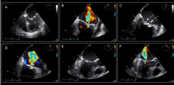
Figure 1. Echocardiographic study.
- Infectious endocarditis Severe mitral.
- Insufficiency secondary to perforation of the posterior leaflet "storm" with vena contract of 6.6 mm
- level Greenery pedicle posterior mitral cusp of 11 × 6 mm diameter
- Auricula dilated left with signs of volume overload
- Pulmonary Hypertension mild psap 40 mmHg
- preserved left ventricular function
- No pericardial effusion in the current study
Personal
Mother-18, father-34, cesarean delivery birth weight-3500 g, Qx: denies, Allergies denies
Physical examination
Ta: 102/46 mmHg Fc: 139, fr 25 SaO2: 94% Weight: 9 kg
Intubated sedated, edematous compatible with noradrenaline milrinone furosemide infusion systolic murmur audible heart sounds in all foci GIII breathing with rales adapted respiratory fan with PMVA 18 10 peep symmetric lung expansion lung expansion symmetrical with mobilizing secretions renal diuresis 483 cc (2.88 cc/kg/hr ) with support diuretic 0.15 mg/kg/hr metabolic alkalosis, metabolic gases suitable electrolytes globose abdominal gastrointestinal depressible no mass or organ enlargement with enteral at 20cc/hr infection : fever in the morning with high procalcitonin linezolid meropenem and amphotericin b hematologic not look pale no bleeding, neurological sedation pinral delayed capillary refill [1].
IDX:
1. Septic shock secondary to cardiogenic more
a. Endocarditis of mitral valve with severe mitral regurgitation
b. More multilobar pneumonia pleural effusion (parapneumonic vs. cardiogenic vs. hiponcótico state)
c. Suspected estafilococcemia (12/12/12 Blood culture gram positive cocci)
d. Septic emboli
2. Ventilatory failure multifactorial
a. Multilobar pneumonia
b. Pleural effusion
c. High suspicion of primary stroke vs septic embolism from endocarditis
3. Multiorgan dysfunction
a. Cardiovascular
b. Respiratory
c. Liver
d. Risk of kidney injury
4. Hiponcótico critical patient status more nutritional crash
Analysis and plan
Patient in critical condition, in cardiogenic shock unwieldy physician demonstrated severe mitral regurgitation due to infectious endocarditis. has indication for surgery to attempt valvuloplasty vs. replacement however it is a complex procedure by the poor condition of the patient and the displayed area of the mitral valve that would hinder the attempt to repair this and limiting with prosthetic valves in children injury.
At the time with positive blood cultures and sirs so in conjunction with PICU choose to continue antibiotic therapy control blood cultures in 3 days to define optimal timing for surgery Also been tried but is compensated heart failure although we know that the probability that the patient gets better from that point of view before surgery is even partially poor [2].
Patient continued deteriorating and considered: given that it may not lead to better clinical status but rather the state is progressively deteriorating, it was decided to advance the surgical procedure.
Findings
Dilated right chambers with signs of pulmonary hypertension, with distension of the pulmonary artery are found. Dilated veins cavas. The mitral valve, with great commitment of the posterior leaflet, with vegetation 2 × 2 cm, with perforation of the entire valve with severe insufficiency, fragility and engagement ring to the back is severely inadequate.
These findings conditional are not able to perform a mitral valve plasty and opt for a change with aortic mechanical prosthesis placed in the mitral position given the cardiac size do meter fitting a ring on aortic -× 21 mm and decided to place an aortic valve mitral invested in position (Figures 2-5).
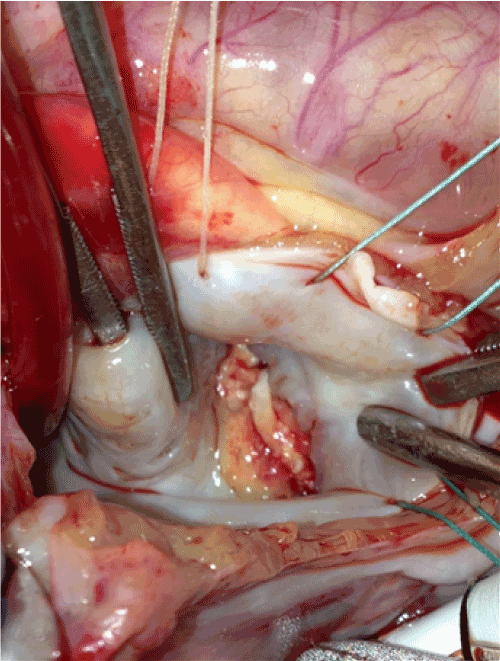
Figure 2. Mitral valve endocarditis.
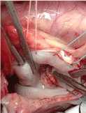
Figure 3. Mitral valve endocarditis
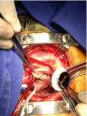
Figure 4. Mitral replacement.
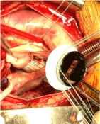
Figure 5. Mitral replacement with aortic mechanical valve.
Good size valve is allowed to avoid reoperation for valve replacement in the short term for placement was in supra- annular position and it was necessary to polish the myocardium to allow proper latch discs.
The pump outlet is uncomplicated sale in sinus rhythm however before you start filtering active bleeding coming from the rear wall and is necessary to enter pump and two small states do not flow for 3 minutes is presented in order to make repair of the posterior wall of the ventricle. there is a defect 3 mm which is related to the site increased friability and commitment of the mitral annulus in the posterior leaflet was found. Once bleeding is controlled repaired and gets out of pump and do the modified filtration.
No bleeding after the procedure is acceptable with left ventricular function and is less distension of the right ventricle and the pulmonary artery.
Immediate Pop
Mechanical ventilation, Milrinone Epinephrine furosemide infusion insulin infusion
TA: 88/61 mmHg, HR 136
Paraclinical
Normal electrolytes, creatinine 0.26 lactic acid 1.2 Gases: Arterial pCO2 ph 7.4 28.5 46 po2 76 sat 95 % HCO3 impaired oxygenation mixed acid-base disorder Venous pH 7.4 PCO2 46 po2 42 sat 77% HCO3 28.5 adequate oxygen extraction delta co2 Normal. It is considered that the development is appropriate, and in the days following his postoperative stabilization of all hemodynamic and respiratory variables was achieved, making the brackets removed. No reactivation of infection or inflammation occurs [3].
Antibiotic therapy is completed, and satisfactory clinical outcome with therapeutic levels of warfarin management decides to give out advice and warning signs you explain to the mother. It verifies that the patient has Warfarin for outpatient management (administered by the early discharge program for the next ten days of therapy) PT and INR monitoring is requested within 3 days and pediatric appointment in 3 days. Patient medication management (Furosemide Enalapril and Spironolactone) mother agrees to get chronic medications.
Cardiovascular surgery
No fever, no dyspnea, recovery of strength in left hemisphere, and gets up and walks a few steps. Eating well
Weight: 9.7 kg
Fc: 100, Fr: 28, good overall condition active mobilized all 4 limbs Heart Sounds with click of valvular rhythmic opening Normal breathing, Abdomen: soft painless Evolution is right, has filed neurological recovery known and nutrition, valve is heard well and has signs of heart failure. Goals in the last inr. At follow-up to date, we have made periodic inspections, and recent develop in the city of Yopal.
We know that the child has adequate evolution with adequate psychomotor development, neurological deficits recovering admission. Take your anticoagulation orderly. His current weight is 18 Kg echocardiography was made two months ago where his mitral valve shows proper operation without parafugas with suitable gradient.
Patients using mechanical valves in our environment have a greater long-term durability, so they are especially useful in young patients. This practice is useful despite the implications of anticoagulation. Particularly in children the benefit in the short and long term shows how alternative for replacement of valves, especially mitral valve, for lack of an alternative graft.
The mitral valve replacement (MVR) and aortic (AVR) is frequent and relatively low morbidity and mortality in adults procedure and imposes special challenges in pediatric patients. It comes with a high surgical morbidity and mortality Among the special considerations that should be focused are the hemodynamic changes related to the small size of the prosthesis and the patient's growth with a relative stenosis of it, is created that the durability of the valve is not defined in this group of patients who have a long life expectancy, which is necessary reoperations in the future, and that patients with mechanical prostheses should receive oral anticoagulation with all the risks and complications involved.
Despite improvements in surgical techniques and the development of better hemodynamic profile prosthesis treatment of valvular disease in children remains controversial and complex valve replacement booking for cases where valve repair is not the choice and the outcome In this case imposed a great challenge to the group, it was possible to obtain an adequate management with a remodeling of the ventricle and the ring to accommodate a valve of an acceptable size for age and thinking about the future growth of the ventricle.
The series described throughout Latin America show an acceptable mortality among pediatric patients being preferred mechanical valve in the mitral position, supranular position. A Chilean series shows in a span of 10 years, treatment of children preferably with mechanical valves, being the most frequent aortic position. These patients require remodeling of the ring. Patients requiring mitral valve were handled in supranular position, and in this group the highest mortality occurred. 17 aortic and 11 mitral valves were operated. The group with the highest mortality was involved with mitral valve replacement. The associated factor was identified in the placement of the prosthesis supranular. The mean age was 6.2 years. These series are similar to those reported in U.S. studies.
In the series of Columbia Presbiteriana a 12% mortality in children shown, an increase in mortality among children under 2 years. The study of Mervyn A. Williams, shows the long- term population treated with ON- X mechanical valves, and handled with a lower level of anticoagulation in Sonia van Riet Provincial Hospital, Port Elizabeth South Africa. Then the data from this important study is the aim of the study was to evaluate the clinical performance of the On- X heart valve in a population of economically disadvantaged students. Most patients were indigenous, low educational level and multiple geographic locations. Between 1999 and 2004, a total of 530 valves (242 mitral valves aortic valves 104 92 double valves) in 438 patients (3-78 years, mean age 33, range) was implanted the most common reason for surgery was rheumatic valve disease (57%), followed by degenerative valve disease (11%) and infectious endocarditis (9%). Follow-up was 95% for a total of 746 patient-years (pt-yr) Among the patient population, 40% were without anticoagulation or anticoagulation unsatisfactorily. Hospital mortality was 2.3% and none of the deaths in the hospital was related to the valve. The mean (± SE) survival (including hospital deaths) at four years was 73.8 ± 8.1% AVR MVR 83.4 ± 5.7 % and 60.9 ± 10.3 % DVR Linearized rates (for AVR, MVR and DVR, respectively) for late complications (%/pTyr ) were: bleeding episodes 0.6, 1.0 and 2.3; thrombosis 0.0, 0.2 and 0.0 ; endocarditis 0.6, 1.0 and 2.3; paravalvular leak 0.6, 0.2, and 0.0; systemic embolism 1.1, 1.5 and 3.5. Most are related to systemic embolism endocarditis. Among the patients there were seven uncomplicated pregnancies to term. Conclusion: Given the erratic anticoagulation coverage and a high incidence of infective endocarditis, results of this study can be considered encouraging. The low incidence of thrombosis valve (one case) was noticeable. These data also suggest that the On-X valve can be implemented with relative safety in women who want children [5].
The enthusiasm after the introduction of mechanical valves during the 1960s was quickly affected by documenting a high incidence of thromboembolic complications. Although the use of anticoagulants reduced the incidence of thrombosis and thromboembolism, the problem of anticoagulant -related hemorrhage is introduced. Consequently, physicians involved in the care of patients with mechanical valves must follow a path between the bleeding with anticoagulants and thrombotic complications Among the large number of mechanical valves have been introduced in clinical practice - all with the hope that the incidence of valve -related complications would be reduced - few have lasted the course and today only a handful are implanted regularly. The two-disc valve ON-X was first used clinically in September 1996 Valve is manufactured from On- X carbon, pyrolytic silicone compound Removing silicon from the manufacturing process results in a much smoother and in theory, a lower surface thrombogenicity The valve housing is of a tubular configuration instead of the configuration of the washer used in other devices The valve has a flared entrance and a natural ratio of length to diameter This design, when combined with the prospects of opening fully resulting in a low linear flow turbulence. These innovative design features suggest that the On-X heart valve.
We want to show this case handled with ON-X valve which would become the youngest patient in the world to be handled with mechanical valve and mitral position obtaining a satisfactory result despite being a method of high mortality.
- Robbins RC, Bowman FO Jr, Malm JR (1988) Cardiac valve replacement in children: a twenty-year series. Ann Thorac Surg 45: 56- 61. [Crossref]
- Peter BR, Patricia FS, Claudio AV, Felipe HR, Gonzalo UM, et al. (2005) Mitral and aortic valve replacement in children: Last decade results with latest generation prothesis. Rev Chil Pediatr 76: 375-383.
- Williams MA, van Riet S (2006) The On-X heart valve: mid-term results in a poorly anticoagulated population. J Heart Valve Dis 15: 80-86. [Crossref]
- Caldarone CA, Raghuveer G, Hills CB, Atkins DL, Burns TL, et al. (2001) Long-term survival after mitral valve replacement in children aged <5 years: a multi-institutional study. Circulation 104: 1143-1147. [Croffref]


