We report such extremely rare case of giant pseudoaneurysm which arises from insertion site of modified Blalock Taussig shunt which was diagnosed by cardiac computed tomography and managed conservatively.
pseudoaneurysm, modified blalock-taussig shunt, cardiac computed tomography
A child with history of tricuspid valve atresia, ventricular septal defect and interrupted aortic arch who underwent initial single ventricle palliation with Norwood procedure, modified Blalock Taussig (BT) shunt placement, and aortic arch repair at age of 11 days. His course was complicated by concerning finding for aneurysm on routine follow up echocardiogram. He had cardiac computed tomography (CT) imaging at age of 2 months. The cardiac CT showed giant aneurysm in the region of modified Blalock Taussig shunt that extends to anterior mediastinum (Figure 1-3). The patient was managed conservatively. At the time of the second palliative surgery, bidirectional Glenn procedure, at age of 7 months, the fibrosed tissue of pseudoaneurysm was resected. Subsequently the patient underwent fenestrated fontan procedure without any complication at age of 3 years and 9 months. He did well after his Fontan procedure.
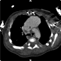
Figure 1A. Cardiac CT reconstructed image in axial view: A: aneurysm, m: proximal modified Blalock Tausig shunt, a: aortic arch
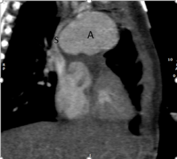
Figure 1B. Cardiac CT reconstructed image in coronal view: A: aneurysm, S: superior vena cava
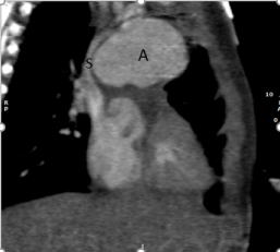
Figure 2A. Cardiac CT reconstructed image in coronal view showing distal modified Blalock Tausig shunt: BT: modified Blalock Taussig shunt, R: Right pulmonary artery
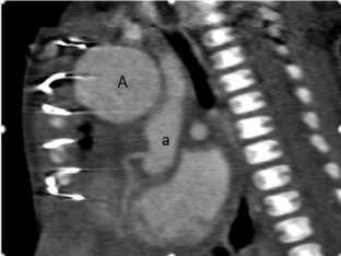
Figure 2B. Cardiac CT reconstructed image in sagittal view showing aneurysm and native aortic root with coronary artery: A: Aneurysm, a: Native aortic root
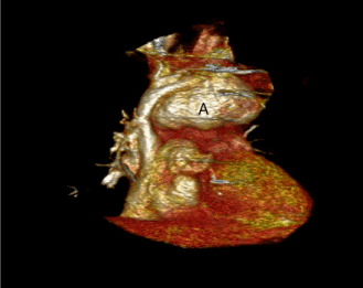
Figure 3A. Cardiac CT Volume rendered reconstruction: Anterior view: A: Aneurysm
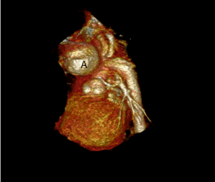
Figure 3B. Cardiac CT Volume rendered reconstruction: Lateral view: A: Aneurysm
Modified BT shunt is commonly performed as the first palliative surgery in single ventricle physiology. Known complications of the modified BT shunt include excessive shunt flow, thrombosis, and pseudoaneurysm [2]. These complications are often managed by emergent surgical intervention with high rates of mortality and morbidity [3]. Here we report very unusual case of giant pseudoaneurysm which arises from insertion site of the shunt diagnosed by Cardiac CT. The patient was managed conservatively and subsequently underwent palliative surgery with good results.
The authors are grateful to the staff of the Cardiovascular computed tomography unit at Children’s Memorial Hermann Hospital for their support in this work.
This research received no specific grant from any funding agency, commercial or non-for-profit sectors.
We know of no conflicts of interest associated with this publication and there has been no financial support for this work that could have influenced its outcome.
According to Children’s Memorial Hermann Hospital Institutional Review Board, this case report was exempt from IRB approval process.
- Parvathy U, Balakrishnan KR, Ranjith MS, Moorthy JS (2002) False aneurysm following
modified Blalock-Taussig shunt. Pediatr Cardiol 23: 178-181. [Crossref]
- Zaki SA, Shanbag P (2010) Pseudoaneurysm following modified Blalock Taussig shunt. Indian Pediatr 47: 198-199. [Crossref]
- Baspinar O, Sahin DA, Sulu A, Gokaslan G (2016) Interventions Involving the Use of
Covered Coronary Artery Stents for Pseudoaneurysms of Blalock-Taussig Shunts. World J Pediatr Congenit Heart Surg 7: 494-497. [Crossref]


