Abstract
Axillofemoral bypass grafts are used to treat symptomatic aorto-iliac vascular disease in patients with comorbidities, that may contraindicate Aortofemoral Bypass (AFB) graft insertion. Most reported cases of AFB graft-associated Pseudoaneurysms (PSA) are at sites of initial anastomoses; in some instances where the initial graft required elongation (thus, new sites of anastomoses), the PSA developed at that location. In contrast, PSA development at the nonanastomatic sites are very rare and usually associated with trauma. We report the first case of a non-traumatic mid-graft PSA in an axillofemoral bypass graft.
Key words
axillofemoral graft, pseudoaneurysm
Introduction
Axillofemoral bypass grafts are used to treat symptomatic aorto-iliac vascular disease in patients with comorbidities, such as those delineated by Slovut et al, that make contraindicate aortofemoral bypass graft insertion [1]. The Axillofemoral Bypass (AFB) is created by instituting a graft of Polytetrafluoroethylene (PTFE) that routes blood flow past a plaque buildup at the iliac artery, anastomosing, most often, at the common femoral artery. AFB is often coupled with a femorofemoral bypass to help supply oxygenated blood to the contralateral lower extremity.
A Pseudoaneurysm (PSA) forms when blood pools in between the two outermost layers of an arterial blood vessel, the tunica media and the strong tunica adventitia. In comparison to a true aneurysm, a PSA excludes involvement of the innermost vessel layer, the tunica media. Only rarely, have there been true aneurysms that develop in AFB grafts [2].
Most reported cases of AFB graft-associated PSAs are at sites of initial anastomoses; in some instances where the initial graft required elongation (thus, new sites of anastomoses), the PSA developed at that location instead [3]. In contrast, PSA development at the nonanastomatic sites has been documented to be very rare and associated with trauma [4].
Case report
The patient is a 71-year-old African American woman who was referred to us for a surgical consultation for worsening bilateral lower extremity claudication and a left-sided mass on her left thorax measuring ~6 cm in diameter. Her past medical history includes long-standing Peripheral Arterial Disease (PAD) controlled by AFB, type II diabetes mellitus, hyperlipidemia, and hypertension. The patient is a current smoker with 10 pack year history.
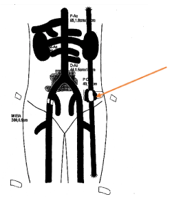
The patient’s initial AFB graft was placed in 2011; Notably, since the initial graft placement in 2011, she had three instances of thrombectomy and revision of the graft: January 2015, March 2016 and, most recently, January of 2018. On physical exam, a large 5-7 cm pulsatile mass was visible on the patient’s left thorax, near the mid axillary line. Vascular exam was positive for a right carotid bruit, non-palpable left femoral pulse, and diminished posterior tibial and dorsalis pedis pulses, bilaterally. Her most recent ABI (July 2018) had shown resting right ABI of 0.34 and a resting left ABI of 1.02. Aorto-iliac duplex examination conducted 1-month prior had measured the mass to measure 2.5 x 2.6 cm with mural thrombus and a residual lumen of 1.2 x 1.3 cm, mass now increased to >6 cm (Figure 1). The patient was afebrile and did not have an elevated WBC count or any other remarkable findings on her physical exam.

Figure 1. A >6cm pseudonauerysm of the graft is identified mid graft
Image courtesy of Ramesh Paladugu, MD, FACS (Texas Vascular and Vein Center)
The patient was taken to surgery for excision of the PSA and a new AFB (re)construction. The patient was placed in the supine position and general anesthesia was implemented. Incisions were made proximally and distally through the skin and the subcutaneous in order to control the flow through the AFB graft; the graft was identified and, then, blood flow controlled. The patient was systemically anticoagulated with 5000 units of heparin and the graft was occluded mechanically with excision to follow later. Before graft excision, we elected to extend an incision over the PSA, disrupting and removing the thrombus within. After clearing out the vascular bed, an 8 mm PTFE graft was placed between the proximal and distal anastomotic sites utilizing 5-0 PROLENE®. We allowed reperfusion before placing surgical glue around the new anastomotic sites. The graft was gently placed underneath the subcutaneous layer, similarly to recorded cases by Orringer et al. [5].
Hemostasis was accomplished by reversing the heparin with protamine sulfate. Wound closure was executed with 0 and 2-0 VICRYL® with intercuticular closure of the wound edges. The patient tolerated the procedure with no complications. She had distal perfusion noted at the completion of the case and the patient was subsequently awakened, extubated, and transferred to the recover area in stable condition. Pathology identified the thrombus material as typical thrombus with organizational change and associated foreign body giant cell reaction; all specimen material excised was negative for malignancy.
Discussion
PSA development is a rare and is usually a result of trauma, either acutely or longstanding, with as evident by the cases documented in review by Mousa et al. [4,6]. Although percutaneous stent placement has been documented in the past by Orringer et al. as an acceptable form of AFB PSA management, we chose to completely replace the graft due to the size of the PSA [5]. In the literature, trauma can be described as a single event (e.g. MVA), a series of a separate events, as with our patient, or it can be over a long periods of chronic irritation, abrasion, and pressure upon the graft [7]. In retrospect, the three instances of thrombectomy and revision of the patient’s graft (January 2015, March 2016 and, most recently, January of 2018) could have warranted a tighter follow-up routine in the patient’s outpatient vascular clinic, which may likely have likely identified the patient’s PSA before it reached the size of ~6cm in diameter. There have been several studies done on the precautionary measures used to take care of AFB graft thrombosis. Notably, Duplex Scan surveillance (DS) may be an affordable and easy-to-execute form of management during routine clinic visits, post-operatively. In their retrospective study on 78 patients, Musicant et al were able to use DS to monitor AFB graft patency and functionality by gathering the Peak Systolic flow Velocities (PSVs); their data showed PSVs < 80 cm/s were associated with increased risks of graft occlusion and sequelae, whereas PSVs > 140 cm/s were a reliable indicator of continued graft patency [8,9].
Conclusion
There are very few documented cases of nonanastamotic PSA development. We present this interesting case in hopes that the findings and the management will contribute to the literature of this rare complication – in doing so, we aim to expedite identification and diagnosis of higher-risk patients, decreasing both morbidity and mortality in this patient population.
References
- Slovut DP, Lipsitz EC (2012) Surgical technique and peripheral artery disease. Circulation 126: 1127-1138. [Crossref]
- Oz BS, Yilmaz AT, Gunay C, Bulakbaayi N, Tatar H (2002) True aneurysm formation in axillofemoral bypass with a reinforced ePTFE graft-a case report. Vasc Endovascular Surg 36: 327-329. [Crossref]
- Jebara V, Aoun T, Dahdah P, Fikani A (2016) Chronic anastomotic pseudoaneurysm on a polytetrafluoroethylene axillobifemoral bypass graft. J Vasc Surg 64: 1484. [Crossref]
2021 Copyright OAT. All rights reserv
- Mousa AY, Nanjundappa A, Abu-Halimah S, Aburahma AF (2013) Traumatic nonanastomotic pseudoaneurysm of axillofemoral bypass graft: a case report and review of the literature. Vasc Endovascular Surg 47: 57-60. [Crossref]
- Orringer MB, Rutherford RB, Skinner DB (1972) An unusual complication of axillofemoral artery bypass. Surgery 72: 769-771. [Crossref]
- Ameli FM, von Schroeder HP, Lossing A (1993) Nonanastomotic pseudoaneurysm of expanded polytetrafluoroethylene axillofemoral bypass graft. J Vasc Surg 17: 777-779. [Crossref]
- Grochow RM, Raffetto JD (2008) Chronic traumatic pseudoaneurysm of polytetrafluoroethylene axillofemoral bypass graft in a quadriplegic patient. Ann Vasc Surg 22: 688-691. [Crossref]
- Musicant SE, Giswold ME, Olson CJ, Landry GJ, Taylor LM Jr, et al. (2003) Postoperative duplex scan surveillance of axillofemoral bypass grafts. J Vasc Surg 37: 54-61. [Crossref]
- Cheng CA, Ho AM (2006) Use of recombinant activated factor VII after axillofemoral bypass grafting. Anaesth Intensive Care 34: 375-358. [Crossref]

