Introduction
The incidence of osteoporotic fractures in industrialized countries is on the rise [1]. The fixation of head fractures of the upper arm (humerus) by percutaneous Kirschner wires (K-wire) or retrograde intramedullary nailing is a simple and quick operative procedure with the advantage of saving the soft tissue and avoiding damage to the vascularization of the treated bone [2,3]. But, this advantages frequently are diminished by the danger of wire loosening with subsequent risk of secondary fracture dislocation. The rate of loosening has been reported between 41% and 73% in the proximal humerus [4]. A previous clinical report by Halder et al. [5] demonstrated the results only of an intramedullary steel nail with a split apex design. The curved wires at the end was supposed to provide proximal fixation and rotation stability. However, dislocations of the wires also occurred with perforation of the calotte.
Another method to reach more stability, are implants made of alloys that can change their shape with temperature. With shape memory alloys (SMAs), devices retain the shape needed for a particular application after deformation above its transformation temperature. They can also be used to improve anchoring of any metallic implants in bone by choosing a shape during the manufacturing which has the potential to increase stability after implantation [6]. Currently, nickel-titanium alloys (Nitinol®, NiTi) are the most useful SMAs due to their high reversible strain property, high superheating tolerance, and low fatigue. Further advantages of NiTi alloys are high corrosion resistance and biocompatibility [7]. Like in orthodontics, angioplasty, and medical engineering, the advantages of devices made from memory alloys have been furthermore established in orthopaedics [8]. Our first results of studies with SMA K-wires applicated in the humerus by drilling through cancellous bone are not practical because of the widening of the drill hole in the bone and the heat during the insertion procedure, causing an early triggering of the memory effect [9]. This preliminary study is conducted in order to evaluate initially only the amount of fixation and rotation resistance of SMA intramedullary nails as parameter of loosening in the bone.
We test now special designed intramedullary nails with split apex made from Nitinol® vs. commercial straight steel nails (Buendel) by comparing the amount of rotation resistance.
Materials and methods
We used SMA-nails with a superelastic memory NiTi alloy with a diameter of 2 mm as raw material (Memory-Metalle Inc., Germany) (Figure 1a). We modified the tip of this nails as a split apex (Figure 1b), manufactured with the use of laser radiation.
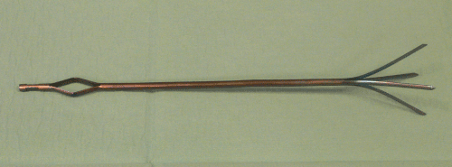
Figure 1a. FGL-nails with NiTi alloy, length 260 mm, diameter 2 mm
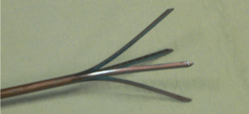
Figure 1b. Detail of the apex of the FGL-nails
The composite ratio was 50% nickel and 50% titanium. The transformation temperature AS was set at 25°C, and wires were stored at –18°C. In the low-temperature phase (martensite) the apex of the SMA-nails could be manually straightened, and then returns to its original shape when released by increasing the temperature above the transformation temperature AS. Mechanical and physical properties were as follows: densitiy, 6, kg/dm³; melting point, 1310 °C; yield stress austenite, 550-700 MPa; youngs modulus of the austenite/martensite, ca. 70-80 46 GPa/ca. 23- 41 GPa depending on the temperature; tensile strain fully annealed/cold worked, 20-60%/ 5-20%.
We used 4 special fixed geminate human humerus bones (8 bones). Distance osteotomy of 5 mm defined at the distal intersection point of the head radius (Figure 2). Measuring of the torsion in Nm/deg with a MTS power test machine with 10 N axial pressure and a torsion of 30°/min. The left humeri were used for the SMA-spreading nails, the right one for the commercial nailing tests. The bone density was on an average 327,55 mg/cm ³, the shaft length of 30,48 cm, the inside diameter 12,7 mm and the head diameter 47,1 mm.
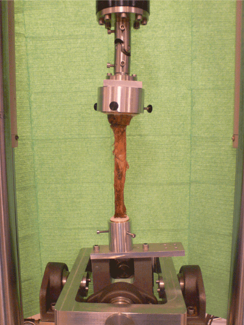
Figure 2. Specimen applied in the test machine.
Results
A worse performance resulted of the SMA nails (0,008325 Nm/deg vs. 0,0014 Nm/deg).
With increased inside diameter of the bone the application of more Bündelnagel was required and the rotation resistance of the SMA nails was worse. A lower Bündelnagel number was required at a decreased inside diameter and a better behavior of the SMA-spreading nails resulted with regard to the rotation (Figure 3-6).
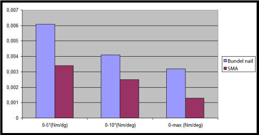
Figure 3. Values of Nm at different degrree of torsion, measured in Nm/deg. Diameter of the bone 19,5 mm
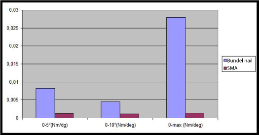
Figure 4. Values of Nm at different degrree of torsion, measured in Nm/deg. Diameter of the bone 18,5 mm
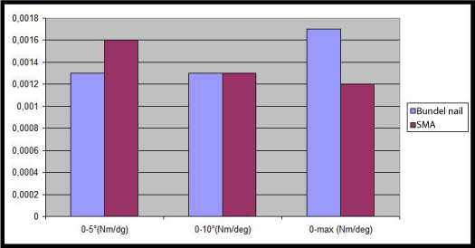
Figure 5. Values of Nm at different degrree of torsion, measured in Nm/deg. Diameter of the bone 14,5 mm
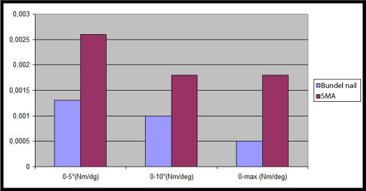
Figure 6. Values of Nm at different degrree of torsion, measured in Nm/deg. Diameter of the bone 12,5 mm
Conclusion
We presume that these first findings are related to the diameter of the medullary cavity. This also applies to the nails. The deadlock in the humerus head seems not to be essential. The inside diameter of the humerus shaft also must be taken into account in the design of the SMA-nail besides the appropriate fracture shape.
References
- Lauritzen JB, Schwarz P, Lund B, McNair P, Transbøl I (1993) Changing incidence and residual lifetime risk of common osteoporosis-related fractures. Osteoporos Int 3: 127-132. [Crossref]
- Brooks CH, Revell WJ, Heatley FW (1993) Vascularity of the humeral head after proximal humeral fractures. An anatomical cadaver study. J Bone Joint Surg Br 75: 132-136. [Crossref]
- Jaberg H, Warner JJ, Jakob RP (1992) Percutaneous stabilization of unstable fractures of the humerus. J Bone Joint Surg Am 74: 508-515. [Crossref]
- Kocialkowski A, Wallace WA (1990) Closed percutaneous K-wire stabilization for displaced fractures of the surgical neck of the humerus. Injury 21: 209-212. [Crossref]
- Halder SC, Chapman JA, Choudhury G, Wallace WA (2001) Retrograde fixation of fractures of the neck and shaft of the humerus with the 'Halder humeral nail'. Injury 32: 695-703. [Crossref]
- Barber FA, Herbert 2021 Copyright OAT. All rights reservength revisited. Arthroscopy 12: 32-38. [Crossref]
- Shabalovskaya SA (1996) On the nature of the biocompatibility and on medical applications of NiTi shape memory and superelastic alloys. Biomed Mater Eng 6: 267-289. [Crossref]
- Haasters J, Sali-Solio G, Bonsman G (1990) The use of Ni-Ti as an implant material in orthopedics. In: Duerig TW, Melton.
- Wiebking U, Gösling T, Monschizada W, Rau T, Krettek C (2007) Do K-wires made from shape memory alloys increase pull-out forces? A preliminary experimental cadaver study in bovine bone. Med Bio Eng Comput 45: 585-589. [Crossref]


