An older of about 11-year-old Labrador retriever dog developed arthrosis especially in the right elbow with a traumatic loosened fragment that was removed in a small operation. The dog was treated first with conventional antiinflammatory drugs, e.g., paracetamol, paracetamol-codeine combination, meloxicam, and finally carprofen-tramadol combination. After these treatments failed to give an adequate response, 40-80 mg methylprednisolone was administered i.m. or s.c. at one to two-month intervals. Skin dermatitis with marked hair loss appeared with clinical appearance of impetigo-like purulent infection seen as multiple lesions. Therefore, cephalexin was administered orally for 4 weeks together with topical antimicrobial treatments with chlorhexidine. In addition, nodular itchy tumors developed. Skin biopsies taken both from areas of dermatitis and nodular lesions revealed marked skin calcification that was treated with oral tetracycline 500 mg bid for 2 months and thereafter 500 mg per day for another 2 months. The itch disappeared in 2 weeks, dermatitis in about 2-3 weeks, and nodular tumors disappeared, and the hair regrew in about 3 months after the end of tetracycline treatment.
skin calcification, tetracycline, treatment
Skin calcification is rather uncommon and can be caused by imbalance of vitamin D and parathyroid hormone (PTH). In humans, skin calcification is seen clearly more seldom than calciphylaxis, that after affecting blood vessels causes skin necrosis and wounds and is often linked to autoimmune diseases as lupus erythematosus, dermatomyositis, scleroderma and CREST syndrome, or malignancies. In addition, calcification can be linked to excess of calcium intake [1-3] and excess of endogenous cortisol production in dogs [4]. Also, iatrogenic corticosteroid can cause skin calcification, especially in large dogs (Labrador retriever, Rottweiler, boxers, Staffordshire terrier), but the steroid dose and other factors causing calcification are poorly known [4].
There are no generally accepted standard therapy for skin calcinosis. However, in humans, several treatments have been reported to be effective mostly in case reports as reviewed by Nadine et al. e.g., warfarin, bisphosphonates, minocycline, ceftriaxone, diltiazem, aluminum hydroxide, probenecid, IVIGs, intralesional corticosteroids, extracorporeal shock wave lithotripsy, surgical intervention and carbon dioxide laser. However, of these, minocycline showed to be effective in 8 of 9 patients, of whom all 8 were positive for antinuclear antibodies and 5 of 8 were positive for anticentromere antibodies [5].
A spayed Pug developed skin calcification when prednisolone was used for the treatment of panniculitis [6]. In the literature, there are no reports on iatrogenic skin calcification in humans. However, in clinical practice we have noticed occasionally skin calcification when high-dose corticosteroids are used for patients having autoimmune diseases, being difficult to evaluate whether calcification is caused by the inflammatory disease or use of corticosteroid or by dual effect of both. In humans, iatrogenic skin calcification has been detected when intravenous calcium-containing solutions are given to patients [7-10]. There is a descriptive report of skin calcinosis in large dogs: German shepherd, Rottweiler and Labrador retriever [11]. Human skin calcinosis has been treated by intravenous sodium thiosulfate [12] and topically by 10% water-oil-emulsion [10] for 6 months, and also, by topically by 25% sodium thiosulfate compounded in zinc oxide in 2 case report [13].
We describe a case report of skin calcification caused by i.m./s.c.- methylprednisolone used for inflammatory arthrosis in a Labrador retriever, and the possible effect of tetracycline to resolve skin calcification.
A yellow Labrador retriever dog was born on June 26, 1999 and the weigth was ca. 37 kg. It gained the title of Finnish Champion in 2002. At the end of 2007 the right elbow joint developed age-related arthrosis treated with oral glucosamines (various products, 270-400 mg daily) and painkillers, first paracetamol (Panadol 500 mg, bid), paracetamol-codeine (Panacod 500mg/30 mg, bid), meloxicam (Metacam 2,5 mg ½ 2-3 times a day) and later carprofen (Canidryl 100 mg ¾-1 bid or Norocarp 50 mg 1½ bid) combined with tramadol (caps Tramadol 50 mg 2 to 3 times a day).
Together with arthrosis, a lock-compulse symptom appeared after a trauma due to a 3-4 mm sized sharp-edged loosen fragment that was surgically removed in October 2009. In basic laboratory tests hemoglobin was normal and alanine aminotransferase, Alat, was 125 U/L (reference 24-136 U/L). Muscle tensions caused by pain due to arthrosis was treated with massage and swimming exercise in a dog pool. In May 2011, an acute hematuria appeared (oxalate crystals and E. coli infection), and in laboratory analysis, Alat (269 U/L) and alkaline fosfatase, Afos (514 U/L, reference 19-106 U/L) were both elevated. Both of these were still elevated 2 weeks later (Alat 438 U/L and Afos 1210 U/L); these abnormalities were interpreted to be caused by used painkillers. After stopping of these drugs, Alat returned to normal (127 U/L) and Afos was almost normal (175 U/L) in 3 months. The diet was changed to Royal Canine Hepatic Dry -food. Urinary infection was treated by amoxicillin-clavulanic acid for 14 days with the dose of 500/125 mg bid. Control urine sample was clean with no crystals.
Right elbow still was painful and swollen, and to reduce the use of painkillers, amitriptyline with the dose of 25 mg bid and oral prednisolone 10 mg /day were administered. After a few weeks, fecal blood was noticed again which was interpreted to be due to oral prednisolone that was stopped. After 2 weeks and ranitidine treatment (150 mg ½ bid) for 3 weeks, fecal blood disappeared. Thereafter, only one oral 10 mg prednisolone 10 mg dose was re-administered followed by re-appearance of fecal blood in 24 hours. Therefore, the treatment was changed to methylprednisolone using 40-80 mg doses i.m. and s.c. at 1 to 2 month intervals. Oral glucosamine was changed to injections with sodium pentosan polysulfate (Cartrophen vet.100 mg/ml s.c.), 4 times in one-week intervals that was later give twice a year.
After about 8 months, hair loss and skin dermatitis developed. Clinically, it was purulent-resembling human impetigo (Figure 1A), and consequently, oral cephalexin and topical chlorhexidine was used. After 4 weeks there was no change in the skin condition, and thus, skin biopsy specimens under local lidocain-adrenaline anesthesia were taken with 4-mm punch from a skin area showing partial hair-loss with diffusely erosive outlook (Figure 1B)and from one representative nodular lesion (Figure 1C). Histologic examination revealed a strong calcification both in the dermatitis area (Figure 2A) and in nodular lesions (Figure 2B).
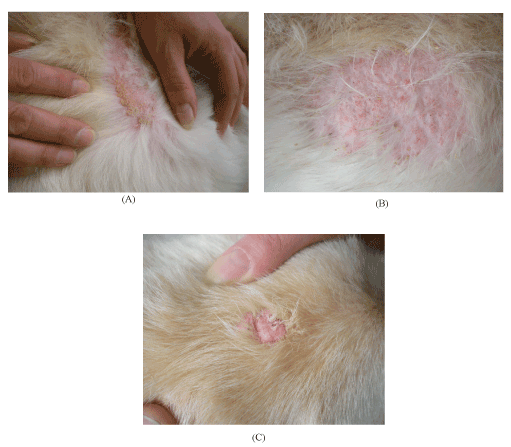
Figure 1. A. Diffuse impetigo-like dermatitis at hair loss area, B. Papuloplaqueous dermatitis at hair loss area, C. Tumorous lesion
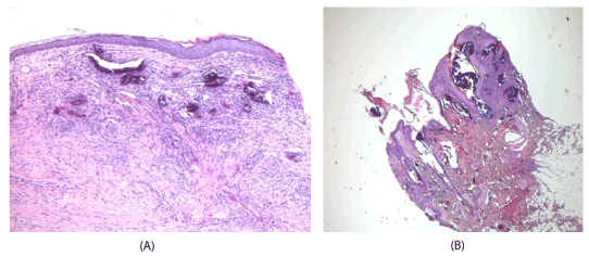
Figure 2. A. Histologic view of dermatitis-hair loss area, B. Histologic view of nodular lesion
In laboratory tests, blood PTH-level was 2,8 pg/ml (reference 4,7 – 54,7 pg/ml) ja Parathormon-related Protein was <0.1 ng/ml (reference<1.0 ng/ml). In cortisol-challenge test, plasma cortisol in the control sample was 98.8 nM and in the 60-min sample 279 nM. Serum calcium was 2.97 mM (reference 2,20-3,10 mM being close the level of upper reference value) and serum phosphate was normal 1,40 mM (reference 0,85-1,95 mM). Other paramaters analyzed were amylase, kreatinine, hemoglobin and blood leucocytes were normal.
By the experience of clinical practice in humans, oral tetracycline was initiated with the dose of 500 mg bid After 2 months, the dose was reduced to 500 mg once a day. Simultaneously a small dose of carprofen (Norocarp vet.) 50mg ½ bid combined with tramadol 50 mg bid) was initiated for 3 weeks. After one month, Alat was 181 U/L and Afos 690 U/L, and additional 6 months later Alat was 167 U/L and Afos was 100 U/L.
After about 3 months of tetracycline initiation the clinical outcome was markedly improved, itch decreased markedly after 2 weeks and disappeared after 3 to 4 weeks together with decreased dermatitis. After about 3 months of the initiation of tetracycline, the clinical outcome was markedly improved. In fact, itch decreased markedly in 2 weeks and disappeared after 3 to 4 weeks together with decreased dermatitis. The hair re-grew more slowly in about 3 months after stopping of tetracycline. Control biopsy specimen was taken close to the same site where the dermatitis biopsy had been taken previously (less than 1 cm away) (Figure 3A). The histology is shown in Figure 3B.
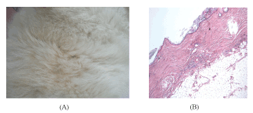
Figure 3. A. Original dermatitis-hair loss area after treatment; complete hair re-grow, B. Original dermatitis-hair loss area (as seen in Figure 3A) after treatment; Histologic view, complete disappearance of calcification
By the experience in humans, the dog received one dose of botulin toxin A intra-articularly (Botox, 40 units/0.4 ml) into the right elbow joint with some clinical pain relief for about one week.
Dog skin calcification is rarely seen at veterinarian’s policlinic and there has been only a few cases in Kuopio Veterinary Center for 20 years. The dog case was challenging to treat since liver enzymes were elevated already in the beginning, and the potent liver toxicity of tetracycline is well known. The dose of tetracycline was selected based on practical clinical experience in dogs and humans. The follow-up of Alat-values revealed that tetracycline dose was moderately tolerated with good clinical treatment response. Skin calcification combined with dermatitis and tumorous changes as well as hair loss completely resolved.
About 2 years after our dog’s successful treatment with tetracycline, a similar case with successful treatment response to minocycline in 2 weeks was reported. In that case, minocycline was given due to iatrogenic calcification after 8-year use of orally administered corticosteroids for allergy in an 8-year-old castrated Schnauzer dog [14]. In the present case, tetracycline was used because minocycline was not available in Finland.
Tetracycline, like also minocycline, are broad-spectrum antibiotics belonging to the tetracycline family. The mechanism of action of tetracyclines on calcinosis may be their property to chelate calcium and directly inhibit many matrix metalloproteinases (MMPs) [15]. These enzymes may be over-expressed in skin calcinosis, and thus, inhibition of these enzymes may play a marked role in reducing inflammation and ulceration [15,16]. Also, tetracyclines are known to decrease calcium salt deposit size [15].
A recent case report of treatment for iatrogenic calcification by topical wash of 90% dimethyl sulfoxide every second day for 2 months showed a good response in a male pug with seropositive leishmaniasis and secondary immune-mediated hemolytic anemia [17]. After treatment of our dog, reports on the treatment of human skin calcinosis by sodium thiosulfate have been published [10,12]. Thus, this might be an alternative option for tetracycline treatment also in dogs. The use of botulin toxin A (Botox) for dog arthrosis is interesting [18], and some response was observed in our dog case. Thus, this treatment may also be an alternative to consider, but economical aspects with limited knowledge on it’s effect were limiting factors in the present case.
It has previously been published that idiopathic and glucocorticoid-associated calcinosis cutis is recognized in English bulldogs, Doberman pinchers, Dachshunds, Rottweilers and Labrador retrievers. The authors report that lesions regress spontaneously within 1 year [19]. Our dog was sick and we decided to get the clinical disease burden lowered faster than waiting for spontaneous relief for months up to one year. Also, the decision was made because of our own clinical experience in humans. In addition, there are published reports on minocycline in the review by Reiter et al. [5] and in the case report by Jang et al. [14]. It is possible that there are variable effects of different members of the tetracycline family, that is, minocycline may possibly give slightly faster response than tetracycline, though the patient number of only 2 dog cases is too small to make conclusions. Therefore, additional case reports are needed to justify clinical trials.
An older Labrador Retriever did not get sufficient treatment response to elbow arthritis by using
conventional (NSAIDs) and potent centrally affecting (tramadol) painkillers, nor was the do without sufficient response to additional treatment with methylprednisolone 40-80 mg i.m. and s.c. at 1-2 month intervals. Instead, the corticosteroid treatment probably was the culprit to induce skin dermatitis, hair loss, nodular lesions and skin calcification
Skin calcification was treated with oral tetracycline; 1,000 mg/day for 2 months followed by 500 mg/day for additional 2 months, with complete resolution. Itch decreased markedly in 2 weeks and disappeared after 3 to 4 weeks together with decreased dermatitis.
We certify that there is no conflict of interest with any financial organization regarding the material discussed in the manuscript.
- Braun-Falco O, Plewig G, Wolff HH, Burgdorf WHC (2000) Chapter 45: Cutaneous calcification: In: Dermatology, 2nd completely revised edition. Berlin: Springer, pp: 1327-1334.
- Reiter N, El-Shabrawi L, Leinweber B, Berghold A, Aberer E (2011) Calcinosis cutis. Part I. Diagnostic pathway. J Am Acad Dermatol 65: 1-12. [Crossref]
- Balin SJ, Wetter DA, Andersen LK, Davis MD (2012) Calcinosis cutis occurring in association with autoimmune connective tissue disease: The mayo clinic experience with 78 patients 1996-2009. Arch Dermatol 148: 455-462. [Crossref]
- Doerr, KA, Outerbridge CA, White SD, Kass, PH, Shiraki R, et al. (2013) Calcinosis cutis in dogs: Histopathological and clinical analysis of 46 cases. Vet Dermatol 24: 355-361-e79. [Crossref]
- Reiter N, El-Shabrawi L, Leinweber B, Berghold A, Aberer E (2011) Calcinosis cutis. Part II. Treatment option. J Am Acad Dermatol 65: 15-22. [Crossref]
- Nakamura M, Kawamura Y, Minegishi M, Momoi Y, Iwasaki T (2004) Hypercalcemia in a dog with resolution of iatrogenic cushing’s syndrome. J Vet Med Sci 66: 329-331. [Crossref]
- Kagen MH, Bansal MG, Grossman M (2000) Calcinosis cutis following the administration of intravenous calcium therapy. Cutis 65: 193-194. [Crossref]
- Hsu CH, Chen WY, Tsai TH (2009) A 9-year-old girl with rhabdomyolysis complicated with iatrogenic calcinosis cutis. Clin Exp Dermatol 34: e503-e504. [Crossref]
- Lim PP, Kossard S, Stapleton K (2012) Calcinosis cutis following contact with calcium chloride solution. Australas J Dermatol 53: e66-e68. [Crossref]
- Garcia-Garcia E, López-López R, Álvarez-Del-Vayo C, Bernabeu-Wittel J (2017) Iatrogenic calcinosis cutis successfully treated with topical sodium thiosulfate. Pediatr Dermatol 34: 356-358. [Crossref]
- Tafti AK, Hanna P, Bourque AC (2005) Calcinosis circumscripta in a dog: A retrospective pathological study. J Vet Med 52: 13-17. [Crossref]
- Chang JJ (2019) Calciphylaxis: Diagnosis, pathogenesis, and treatment. Adv Skin Wound Care 32: 205-215. [Crossref]
- Bair B, Fivenson D (2011) A novel treatment for ulcerative calcinosis cutis. J Drugs Dermatol 10: 1042-1044. [Crossref]
- Jang HJ, Kang MH, Sur JH, Park HM (2013) Minocycline as a treatment of dog with calcinosis cutis. Korean J Vet Res 53: 253-256.
- Robertson LP, Marshall RW, Hickling P (2003) Treatment of cutaneous calcinosis in limited systemic sclerosis with minocycline. Ann Rheum Dis 62: 267-269. [Crossref]
- Sapadin AN, Fleischmajer R (2006) Tetracyclines: Nonantibiotic properties and their clinical implications. J Am Acad Dermatol 54: 258-265.[Crossref]
- Tolon JMC, Jiminez JJE, Irizar IG, Trasobares PC (2018) Resolution of iatrogenic calcinosis cutis in a dog through topical application of DMSO. Vet Rec Case Rep 6: 1-2.
- Nicácio GM, Luna SPL, Cavaleti P, Cassu RN (2019) Intra-articular botulinum toxin A (BoNT/A) for pain management in dogs with osteoarthritis secondary to hip dysplasia: A randomized controlled clinical trial. J Vet Med Sci 81: 411-417. [Crossref]
- Scott DW, Miller WH Jr, Griffin CE, Eds. (2001) Chapter 20, Calcinosis cutis. In: Muller & Kirk’s Small Animal Dermatology, 6th edition, Philadelphia: 1398-1399.